Copyright
©The Author(s) 2016.
World J Gastroenterol. Jun 21, 2016; 22(23): 5342-5352
Published online Jun 21, 2016. doi: 10.3748/wjg.v22.i23.5342
Published online Jun 21, 2016. doi: 10.3748/wjg.v22.i23.5342
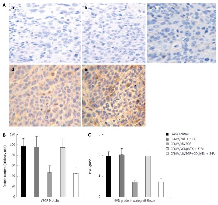
Figure 6 Immunohistochemistry analysis for yCDglyTK and vascular endothelial growth factor in EC9706 xenograft sections.
A: Histological expression and distribution of yCDglyTK at magnification × 200; no-treatment control group (a); CPNPs/null + 5-FC (b); CPNPs/shVEGFX (c); CPNPs/yCDglyTK+5-FC (d); CPNPs/shVEGF-yCDglyTK +5-FC (e); B: Integrated optical density (IOD) values of VEGF expression in EC9706 xenografts. Anti-VEGF antibody was used for immunohistochemistry assay; C: Quantification of angiogenesis by microvessel counts (MVC) in EC9706 xenografts. Anti-CD34 was used for microvessel staining. CPNPs: Calcium phosphate nanoparticles; VEGF: Vascular endothelial growth factor; 5-FC: 5-fluorocytosine.
- Citation: Liu T, Wu HJ, Liang Y, Liang XJ, Huang HC, Zhao YZ, Liao QC, Chen YQ, Leng AM, Yuan WJ, Zhang GY, Peng J, Chen YH. Tumor-specific expression of shVEGF and suicide gene as a novel strategy for esophageal cancer therapy. World J Gastroenterol 2016; 22(23): 5342-5352
- URL: https://www.wjgnet.com/1007-9327/full/v22/i23/5342.htm
- DOI: https://dx.doi.org/10.3748/wjg.v22.i23.5342


