Copyright
©The Author(s) 2016.
World J Gastroenterol. Jun 21, 2016; 22(23): 5342-5352
Published online Jun 21, 2016. doi: 10.3748/wjg.v22.i23.5342
Published online Jun 21, 2016. doi: 10.3748/wjg.v22.i23.5342
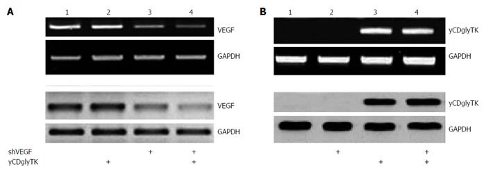
Figure 3 Changes of vascular endothelial growth factor and yCDglyTK expression in established stable cell lines.
A: Representative VEGF mRNA and protein expression were analyzed by RT-PCR (top panel) and western blot (bottom panel), respectively. GAPDH was used as an internal control. Lane 1, EC9706/null; lane 2, EC9706/yCDglyTK; lane 3, EC9706/shVEGF; lane 4, EC9706/shVEGF-yCDglyTK; B: Representative yCDglyTK mRNA and protein expression were analyzed by semiquantitative RT-PCR (top panel) and western blot (bottom panel), respectively. GAPDH was used as an internal control. Lane 1, EC9706/null; lane 2, EC9706/shVEGF; lane 3, EC9706/yCDglyTK; lane 4, EC9706/shVEGF-yCDglyTK. GAPDH: Glyceraldehyde-3-phosphate dehydrogenase; RT-PCR: Reverse transcription polymerase chain reaction; VEGF: Vascular endothelial growth factor.
- Citation: Liu T, Wu HJ, Liang Y, Liang XJ, Huang HC, Zhao YZ, Liao QC, Chen YQ, Leng AM, Yuan WJ, Zhang GY, Peng J, Chen YH. Tumor-specific expression of shVEGF and suicide gene as a novel strategy for esophageal cancer therapy. World J Gastroenterol 2016; 22(23): 5342-5352
- URL: https://www.wjgnet.com/1007-9327/full/v22/i23/5342.htm
- DOI: https://dx.doi.org/10.3748/wjg.v22.i23.5342


