Copyright
©The Author(s) 2016.
World J Gastroenterol. Jun 14, 2016; 22(22): 5297-5300
Published online Jun 14, 2016. doi: 10.3748/wjg.v22.i22.5297
Published online Jun 14, 2016. doi: 10.3748/wjg.v22.i22.5297
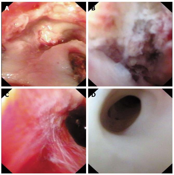
Figure 2 Endoscopic images of portal vein before and after thrombectomy.
A: Before thrombectomy, endoscopy revealed scattered tissue of tumor thrombus near the opening stump; B: Residual tumor thrombus was adhered to the inner wall of the portal vein near the conjunction; C: After repeated retraction of the residual tumor thrombus, endoscopy revealed a clean inner wall of the portal vein with no macroscopic thrombus remaining; D: The left secondary branch of the portal vein was clean with no scattered thrombus.
- Citation: Li N, Wei XB, Cheng SQ. Application of cystoscope in surgical treatment of hepatocellular carcinoma with portal vein tumor thrombus. World J Gastroenterol 2016; 22(22): 5297-5300
- URL: https://www.wjgnet.com/1007-9327/full/v22/i22/5297.htm
- DOI: https://dx.doi.org/10.3748/wjg.v22.i22.5297


