Copyright
©The Author(s) 2016.
World J Gastroenterol. Jun 14, 2016; 22(22): 5154-5164
Published online Jun 14, 2016. doi: 10.3748/wjg.v22.i22.5154
Published online Jun 14, 2016. doi: 10.3748/wjg.v22.i22.5154
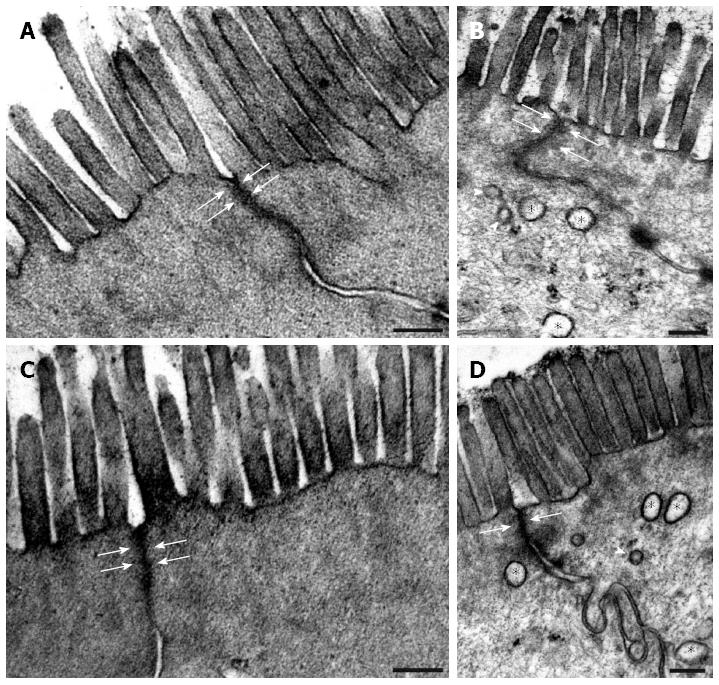
Figure 3 Representative electron micrographs of two neighbouring enterocytes from the colon of control animals (A, C) and in rats treated three times with 2,4,6-trinitrobenzenesulfonic acid.
On days 90 (B) and 120 (D) following the third 2,4,6-trinitrobenzenesulfonic acid (TNBS) administration, both the microvillar surface and the width of the apical intercellular tight junctions (arrows) of the enterocytes in the strictured regions were similar to those in the age-matched controls (A on day 90 and C on day 120). However, autophagosomes (asterisks) and lysosomes (arrowheads) were commonly observed within the epithelial cells of the strictured gut wall. Bars: 200 nm.
- Citation: Talapka P, Berkó A, Nagy LI, Chandrakumar L, Bagyánszki M, Puskás LG, Fekete &, Bódi N. Structural and molecular features of intestinal strictures in rats with Crohn's-like disease. World J Gastroenterol 2016; 22(22): 5154-5164
- URL: https://www.wjgnet.com/1007-9327/full/v22/i22/5154.htm
- DOI: https://dx.doi.org/10.3748/wjg.v22.i22.5154


