Copyright
©The Author(s) 2016.
World J Gastroenterol. Jun 7, 2016; 22(21): 4988-4998
Published online Jun 7, 2016. doi: 10.3748/wjg.v22.i21.4988
Published online Jun 7, 2016. doi: 10.3748/wjg.v22.i21.4988
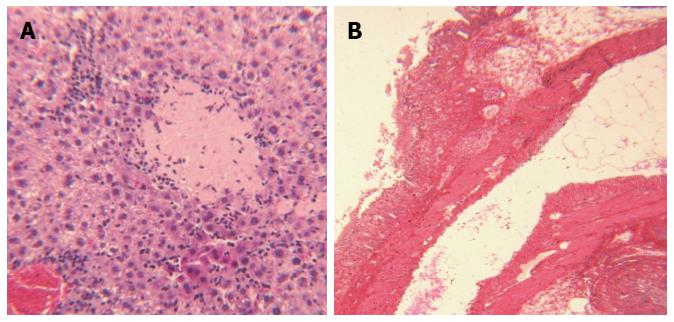
Figure 7 Photomicrograph of hepatitis (A) and colitis (B) from dibutyltin dichloride-treated animals.
A: Hepatitis: Photomicrograph illustrates necrosis of hepatocytes, infiltration of inflammatory cells (magnification × 40). Representative slide from n = 8 animals/group; B: Colitis: Photomicrograph demonstrates distortion of colonic microstructure, loss of brush boarder epithelial cells, infiltration of inflammatory cells into mucosa and cryptic abscess formation. Representative slide from n = 8 animals/group.
- Citation: Oz HS. Multiorgan chronic inflammatory hepatobiliary pancreatic murine model deficient in tumor necrosis factor receptors 1 and 2. World J Gastroenterol 2016; 22(21): 4988-4998
- URL: https://www.wjgnet.com/1007-9327/full/v22/i21/4988.htm
- DOI: https://dx.doi.org/10.3748/wjg.v22.i21.4988


