Copyright
©The Author(s) 2016.
World J Gastroenterol. May 28, 2016; 22(20): 4891-4900
Published online May 28, 2016. doi: 10.3748/wjg.v22.i20.4891
Published online May 28, 2016. doi: 10.3748/wjg.v22.i20.4891
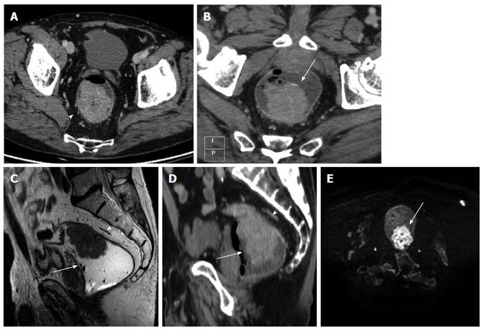
Figure 1 Images obtained in a 54 years-old man with middle-high rectal cancer.
A: The pure axial contrast enhanced computed tomography (CE-CT) image shows a tumor, as a intraluminal polypoid mass, with spiculated configuration margin and spread through the mesorectal fat. The tumour does not involve the mesorectal fascia (MRF) (arrowhead); B: Multiplanar reconstruction (MPR) MDCT images, along the axial plane of tumour axis, shows the presence of the tumor (arrow) with no involvement of the MRF; C: T2 (TSE) MRI image of the same patient (sagittal plane), shows the tumor as a polypoid mass (arrow), along the posterior burden of the rectum infiltrating through the muscularis propria into the mesorectal fat without MRF involvement (arrowhead); D: CE-CT MPR image (sagittal slice), at the same level shows the tumour as a polypoid mass inside the rectal lumen infiltrating the mesorectal fat without MRF involvement (arrowhead); E: DWIBS image (b-value 1000), the tumor presents high signal, due to restricted water diffusion; the normal rectal wall or the surrounding tissues have a lower signal intensity in comparison with the polypoid mass. The mesorectal (arrow) fascia is not detectable.
- Citation: Ippolito D, Drago SG, Franzesi CT, Fior D, Sironi S. Rectal cancer staging: Multidetector-row computed tomography diagnostic accuracy in assessment of mesorectal fascia invasion. World J Gastroenterol 2016; 22(20): 4891-4900
- URL: https://www.wjgnet.com/1007-9327/full/v22/i20/4891.htm
- DOI: https://dx.doi.org/10.3748/wjg.v22.i20.4891


