Copyright
©The Author(s) 2016.
World J Gastroenterol. May 28, 2016; 22(20): 4835-4847
Published online May 28, 2016. doi: 10.3748/wjg.v22.i20.4835
Published online May 28, 2016. doi: 10.3748/wjg.v22.i20.4835
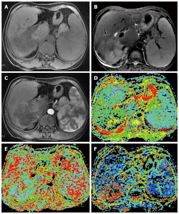
Figure 2 Fifty-three-year-old female patient with hepatocellular carcinoma in the right lobe of the liver.
A: Axial T1-weighted image shows a hypointense mass lesion; B: Axial T2-weighted image shows a hyperintense mass lesion; C: Contrast-enhanced MRI during the arterial phase showing lesion enhancement; D: Mapping of the estimated value of the D parameter. The average value in the lesion ROI was D = 1.22 × 10⁻³ mm2/s; E: Mapping of the estimated value of the D* parameter. The average value in the lesion ROI was D* = 20.6 × 10⁻³ mm2/s; F: Mapping of the perfusion fraction (f) with a value of 19.6%.
- Citation: Yang K, Zhang XM, Yang L, Xu H, Peng J. Advanced imaging techniques in the therapeutic response of transarterial chemoembolization for hepatocellular carcinoma. World J Gastroenterol 2016; 22(20): 4835-4847
- URL: https://www.wjgnet.com/1007-9327/full/v22/i20/4835.htm
- DOI: https://dx.doi.org/10.3748/wjg.v22.i20.4835


