Copyright
©The Author(s) 2016.
World J Gastroenterol. Jan 14, 2016; 22(2): 853-861
Published online Jan 14, 2016. doi: 10.3748/wjg.v22.i2.853
Published online Jan 14, 2016. doi: 10.3748/wjg.v22.i2.853
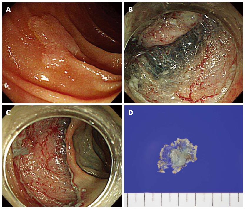
Figure 3 Endoscopic submucosal dissection.
A: A 12 mm sized superficial elevated type (IIa) lesion in the second portion of the duodenum; B: Circumferential mucosal incision and submucosal dissedtion; C: The lesion successfully removed en bloc without complications; D: A 27 mm resected specimen with adenoma.
- Citation: Lim CH, Cho YS. Nonampullary duodenal adenoma: Current understanding of its diagnosis, pathogenesis, and clinical management. World J Gastroenterol 2016; 22(2): 853-861
- URL: https://www.wjgnet.com/1007-9327/full/v22/i2/853.htm
- DOI: https://dx.doi.org/10.3748/wjg.v22.i2.853


