Copyright
©The Author(s) 2016.
World J Gastroenterol. Jan 14, 2016; 22(2): 727-735
Published online Jan 14, 2016. doi: 10.3748/wjg.v22.i2.727
Published online Jan 14, 2016. doi: 10.3748/wjg.v22.i2.727
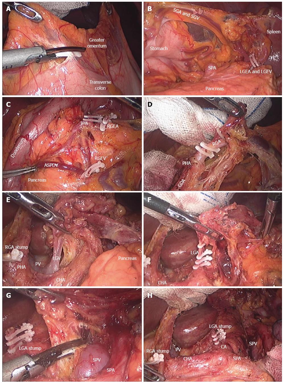
Figure 1 Intraoperative view during distal subtotal gastrectomy with D2 lymph node dissection.
A: Division of the greater omentum; B: Isolation of the LGEA and LGEV; C: Exposure of the RGEA, RGEV, and ASPDV; D: Isolation of the RGA; E: Dissection of the hepatoduodenal ligament, exposure of the PV and isolation of the LGV; F: Isolation and ligation of the LGA; G: Dissection along the SPA and SPV; H: Suprapancreatic view after D2 lymph node dissection. RGEA: Right gastroepiploic artery; LGEA: Left gastroepiploic artery; LGEV: Left gastroepiploic vein; RGEV: Right gastroepiploic vein; ASPDV: Anterior superior pancreaticoduodenal vein; RGA: Right gastric artery; LGA: Left gastric artery; SPA: Splenic artery; SPV: Splenic vein; CHA: Common hepatic artery; GDA: Gastroduodenal artery; IPA: Infrapyloric artery; LGV: Left gastric vein; PHA: Proper hepatic artery; PV: Portal vein; SGA: Short gastric artery; SGV: Short gastric vein.
- Citation: Son T, Hyung WJ. Laparoscopic gastric cancer surgery: Current evidence and future perspectives. World J Gastroenterol 2016; 22(2): 727-735
- URL: https://www.wjgnet.com/1007-9327/full/v22/i2/727.htm
- DOI: https://dx.doi.org/10.3748/wjg.v22.i2.727


