Copyright
©The Author(s) 2016.
World J Gastroenterol. Jan 14, 2016; 22(2): 600-617
Published online Jan 14, 2016. doi: 10.3748/wjg.v22.i2.600
Published online Jan 14, 2016. doi: 10.3748/wjg.v22.i2.600
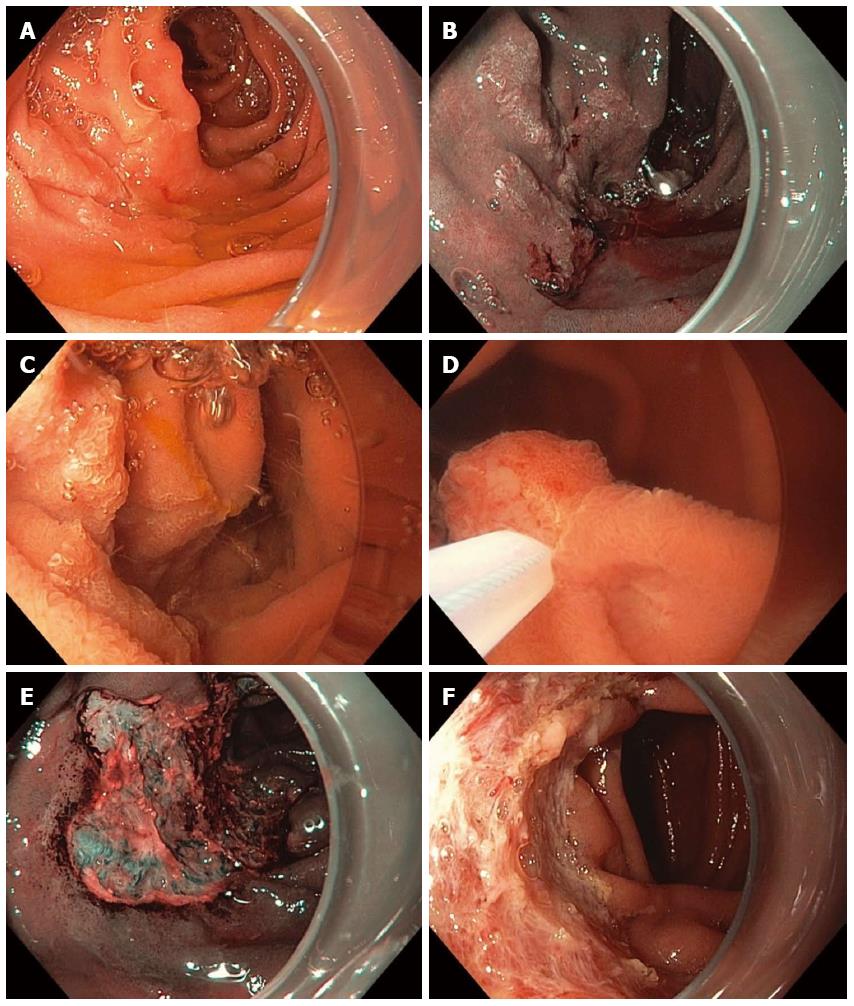
Figure 11 Piecemeal underwater endoscopic mucosal resection.
A large, 3-cm, non-ampullary, laterally spreading adenoma was found in the second portion of the duodenum (A); Narrow-band imaging (NBI) demonstrated adenoma without evidence of invasive cancer (B); Carbon dioxide gas that was used for insufflation was removed and water infused (C); A 15-mm crescent snare (D) was used for piecemeal underwater endoscopic mucosal resection (UEMR). The electrosurgical settings used for snare resection were DRYCUT 60 W, Effect 4, using a VIO 300D generator (ERBE USA, Marietta, GA). NBI was used to identify a residual island of adenoma (E, seen just inside the “9 o’clock” margin of the resection). Final images showed complete endoscopic resection (F).
- Citation: Gaspar JP, Stelow EB, Wang AY. Approach to the endoscopic resection of duodenal lesions. World J Gastroenterol 2016; 22(2): 600-617
- URL: https://www.wjgnet.com/1007-9327/full/v22/i2/600.htm
- DOI: https://dx.doi.org/10.3748/wjg.v22.i2.600


