Copyright
©The Author(s) 2016.
World J Gastroenterol. Jan 14, 2016; 22(2): 546-556
Published online Jan 14, 2016. doi: 10.3748/wjg.v22.i2.546
Published online Jan 14, 2016. doi: 10.3748/wjg.v22.i2.546
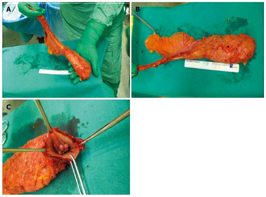
Figure 4 Surgical specimen of robot-assisted total mesorectal excision.
A: This figure shows a very smooth mesorectal fat pad which tapers distally consistent with complete excision of the mesorectum. Grade 3 according to Quirke; B: Grade 3 complete excision according to Quirke showing the wisps of fascia that surrounds the mesorectum indicating that a fascial plane actually exists and points to a complete excision by surgeon, mesorectal vessels contained within the mesorectum can be seen; arrow head; C: Opened specimen showing the tumor; arrow head.
- Citation: Biffi R, Luca F, Bianchi PP, Cenciarelli S, Petz W, Monsellato I, Valvo M, Cossu ML, Ghezzi TL, Shmaissany K. Dealing with robot-assisted surgery for rectal cancer: Current status and perspectives. World J Gastroenterol 2016; 22(2): 546-556
- URL: https://www.wjgnet.com/1007-9327/full/v22/i2/546.htm
- DOI: https://dx.doi.org/10.3748/wjg.v22.i2.546


