Copyright
©The Author(s) 2016.
World J Gastroenterol. May 21, 2016; 22(19): 4776-4780
Published online May 21, 2016. doi: 10.3748/wjg.v22.i19.4776
Published online May 21, 2016. doi: 10.3748/wjg.v22.i19.4776
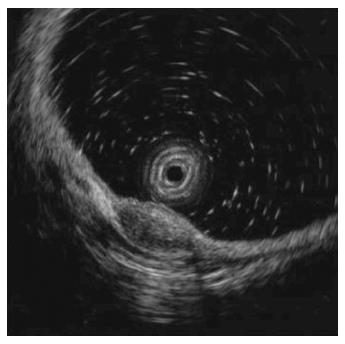
Figure 2 Endoscopic ultrasonographic features of the rectal submucosal tumor.
An approximately 1.0 cm × 0.5 cm, hypoechoic lesion in the deep mucosa and submucosal layer.
- Citation: Kim JK, Baek DH, Lee BE, Kim GH, Song GA, Park DY. Endoscopic resection of sparganosis presenting as colon submucosal tumor: A case report. World J Gastroenterol 2016; 22(19): 4776-4780
- URL: https://www.wjgnet.com/1007-9327/full/v22/i19/4776.htm
- DOI: https://dx.doi.org/10.3748/wjg.v22.i19.4776


