Copyright
©The Author(s) 2016.
World J Gastroenterol. May 21, 2016; 22(19): 4741-4749
Published online May 21, 2016. doi: 10.3748/wjg.v22.i19.4741
Published online May 21, 2016. doi: 10.3748/wjg.v22.i19.4741
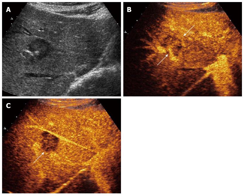
Figure 4 Contrast-enhanced ultrasound feature of hepatic epithelioid hemangioendothelioma in right lobe of liver.
A: Grayscale ultrasound showed a hypoechoic focal liver lesions (FLL); B: In the arterial phase the lesion showed heterogeneous enhancement (22 s after injection of SonoVue); C: In the portal venous phase (53 s after injection of SonoVue), the lesion washed out fast and showed hypoenhancement.
- Citation: Dong Y, Wang WP, Cantisani V, D’Onofrio M, Ignee A, Mulazzani L, Saftoiu A, Sparchez Z, Sporea I, Dietrich CF. Contrast-enhanced ultrasound of histologically proven hepatic epithelioid hemangioendothelioma. World J Gastroenterol 2016; 22(19): 4741-4749
- URL: https://www.wjgnet.com/1007-9327/full/v22/i19/4741.htm
- DOI: https://dx.doi.org/10.3748/wjg.v22.i19.4741


