Copyright
©The Author(s) 2016.
World J Gastroenterol. May 21, 2016; 22(19): 4685-4694
Published online May 21, 2016. doi: 10.3748/wjg.v22.i19.4685
Published online May 21, 2016. doi: 10.3748/wjg.v22.i19.4685
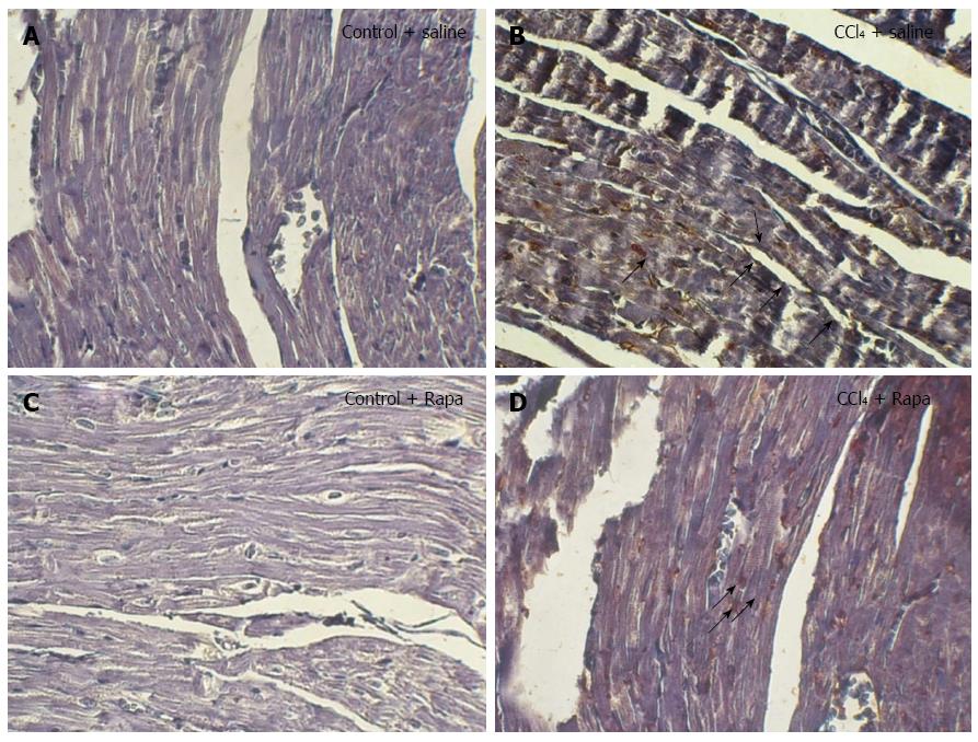
Figure 6 Immunohistochemical staining for p-mTOR in the ventricles of the rats in the following groups: control, cirrhotic, control + rapamycin and cirrhotic + rapamycin (× 400 magnification).
Human gastric tissue was used as the positive control. Note the increased immunostaining of p-mTOR in the myocytes of the rats with cirrhosis. No significant immunostaining was localized to the cardiomyocytes of the untreated cirrhotic rats. In contrast, treatment with rapamycin caused significant immunostaining in the cardiomyocytes of the cirrhotic rats. The black arrows indicate to the p-mTOR immunoblots in rat ventricles.
- Citation: Saeedi Saravi SS, Ghazi-Khansari M, Ejtemaei Mehr S, Nobakht M, Mousavi SE, Dehpour AR. Contribution of mammalian target of rapamycin in the pathophysiology of cirrhotic cardiomyopathy. World J Gastroenterol 2016; 22(19): 4685-4694
- URL: https://www.wjgnet.com/1007-9327/full/v22/i19/4685.htm
- DOI: https://dx.doi.org/10.3748/wjg.v22.i19.4685


