Copyright
©The Author(s) 2016.
World J Gastroenterol. May 21, 2016; 22(19): 4626-4637
Published online May 21, 2016. doi: 10.3748/wjg.v22.i19.4626
Published online May 21, 2016. doi: 10.3748/wjg.v22.i19.4626
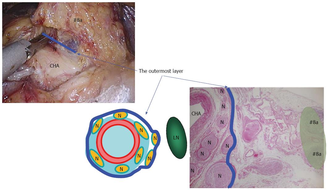
Figure 1 Outermost layer of the autonomic nerve.
Shown in the blue line, lies between the vascular sheath of the major arteries and the fat tissue including lymph nodes. Appropriate tension given to this thin loose connective tissue layer generates sufficient space for safe, adequate and reproducible prophylactic lymph node dissection along the major arteries. LN: Lymph node; N: Nerve; CHA: Common hepatic artery.
- Citation: Suda K, Nakauchi M, Inaba K, Ishida Y, Uyama I. Minimally invasive surgery for upper gastrointestinal cancer: Our experience and review of the literature. World J Gastroenterol 2016; 22(19): 4626-4637
- URL: https://www.wjgnet.com/1007-9327/full/v22/i19/4626.htm
- DOI: https://dx.doi.org/10.3748/wjg.v22.i19.4626


