Copyright
©The Author(s) 2016.
World J Gastroenterol. May 14, 2016; 22(18): 4567-4575
Published online May 14, 2016. doi: 10.3748/wjg.v22.i18.4567
Published online May 14, 2016. doi: 10.3748/wjg.v22.i18.4567
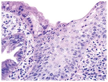
Figure 2 Histological section of gastric-type columnar mucosa of the esophagus.
Moderate mononuclear infiltrate in the lamina propria and polymorphonuclear leukocytes infiltrating the mucosa layer can be observed (Hematoxylin-eosin staining stain; original magnification × 40).
- Citation: Herrera-Goepfert R, Oñate-Ocaña LF, Mosqueda-Vargas JL, Herrera LA, Castro C, Mendoza J, González-Barrios R. Methylation of DAPK and THBS1 genes in esophageal gastric-type columnar metaplasia. World J Gastroenterol 2016; 22(18): 4567-4575
- URL: https://www.wjgnet.com/1007-9327/full/v22/i18/4567.htm
- DOI: https://dx.doi.org/10.3748/wjg.v22.i18.4567


