Copyright
©The Author(s) 2016.
World J Gastroenterol. May 14, 2016; 22(18): 4466-4483
Published online May 14, 2016. doi: 10.3748/wjg.v22.i18.4466
Published online May 14, 2016. doi: 10.3748/wjg.v22.i18.4466
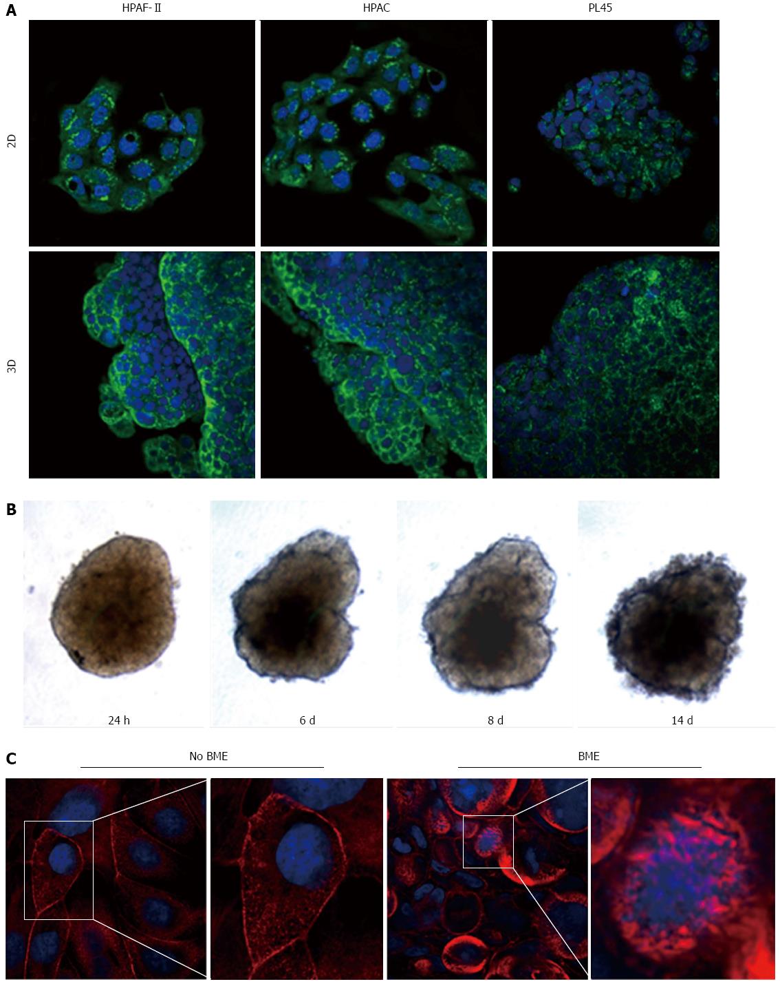
Figure 9 Podoplanin expression and actin cytoskeleton.
A: Micrographs with a confocal microscope of PDAC cells grown in 2D-monolayers and 3D-spheroids, showing the expression of podoplanin. Punctuate immunoreactivity is located in the cytoplasm. Original magnification: 60 ×; B: HPAF-II 3D-spheroid grown in basement membrane extract (BME) monitored at different time points; C: HPAF-II 3D-spheroids grown in the absence or presence of BME were stained using rhodamine-phalloidin to detect actin filaments. Invadopodia are evident boxed. Original magnification: 60 ×.
- Citation: Gagliano N, Celesti G, Tacchini L, Pluchino S, Sforza C, Rasile M, Valerio V, Laghi L, Conte V, Procacci P. Epithelial-to-mesenchymal transition in pancreatic ductal adenocarcinoma: Characterization in a 3D-cell culture model. World J Gastroenterol 2016; 22(18): 4466-4483
- URL: https://www.wjgnet.com/1007-9327/full/v22/i18/4466.htm
- DOI: https://dx.doi.org/10.3748/wjg.v22.i18.4466


