Copyright
©The Author(s) 2016.
World J Gastroenterol. May 14, 2016; 22(18): 4466-4483
Published online May 14, 2016. doi: 10.3748/wjg.v22.i18.4466
Published online May 14, 2016. doi: 10.3748/wjg.v22.i18.4466
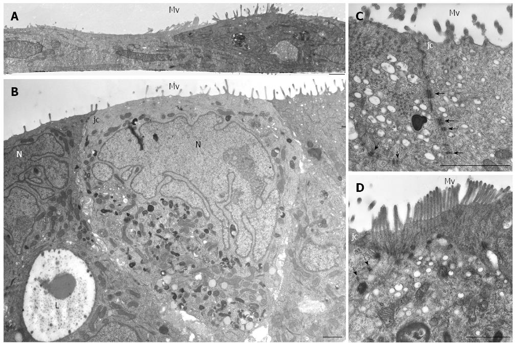
Figure 2 HPAF-II ultrastructure.
A: Electron microscopy of HPAF-II culture grown in a 2D-monolayer showing two cells partially overlapping and exhibiting microvilli (Mv). Scale bar = 1 μm; B-D: Ultrastructural features of HPAF-II cells grown in 3D-spheroids. Adjacent cells located at the periphery of the spheroid show microvilli (Mv) in the apical domain and junctional complexes (jc). In B, some cells exhibit a pale and markedly irregular nucleus (N). Some adjacent cells delimit a lumen-like structure (L). Scale bar = 1.5 μm. C, D: Adjoined cells are connected by junctional complexes (jc) in their apical-lateral domains and by numerous adherens junctions (arrows) in their lateral domains. In D, microvilli are densely packed; at their core, actin microfilaments can be seen extending into the apical cytoplasm. C, D: scale bar = 1.5 μm.
- Citation: Gagliano N, Celesti G, Tacchini L, Pluchino S, Sforza C, Rasile M, Valerio V, Laghi L, Conte V, Procacci P. Epithelial-to-mesenchymal transition in pancreatic ductal adenocarcinoma: Characterization in a 3D-cell culture model. World J Gastroenterol 2016; 22(18): 4466-4483
- URL: https://www.wjgnet.com/1007-9327/full/v22/i18/4466.htm
- DOI: https://dx.doi.org/10.3748/wjg.v22.i18.4466


