Copyright
©The Author(s) 2016.
World J Gastroenterol. May 7, 2016; 22(17): 4421-4426
Published online May 7, 2016. doi: 10.3748/wjg.v22.i17.4421
Published online May 7, 2016. doi: 10.3748/wjg.v22.i17.4421
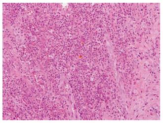
Figure 3 Numerous acute inflammatory cell and limphoplasma cell surrounding fibrosis and granulation tissue is seen.
Bile stained gallstone sludge is seen in the center (haematoxylin and eosin staining, original magnification × 200).
- Citation: Kim BS, Joo SH, Kim HC. Spilled gallstones mimicking a retroperitoneal sarcoma following laparoscopic cholecystectomy. World J Gastroenterol 2016; 22(17): 4421-4426
- URL: https://www.wjgnet.com/1007-9327/full/v22/i17/4421.htm
- DOI: https://dx.doi.org/10.3748/wjg.v22.i17.4421


