Copyright
©The Author(s) 2016.
World J Gastroenterol. Apr 28, 2016; 22(16): 4259-4263
Published online Apr 28, 2016. doi: 10.3748/wjg.v22.i16.4259
Published online Apr 28, 2016. doi: 10.3748/wjg.v22.i16.4259
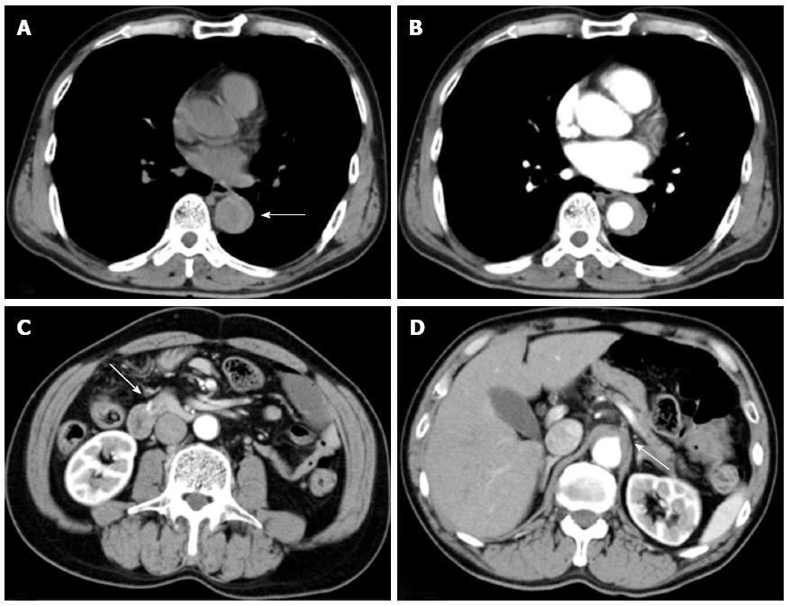
Figure 1 Initial computed tomography performed at local hospital before the onset of hypotension.
A: Plain computed tomography (CT) demonstrating dilated descending aorta and hyperattenuated collection located eccentrically within the aortic wall (arrow); B: Contrast-enhanced CT revealed crescentic and asymmetric wall thickening of the descending aorta: Contrast is not visualized within the aortic media. Intimal flap was not detected; C: An aneurysm at the anteroinferior pancreaticoduodenal artery (arrow); D: The occluded root of the coeliac artery (arrow).
- Citation: Sakatani A, Doi Y, Kitayama T, Matsuda T, Sasai Y, Nishida N, Sakamoto M, Uenoyama N, Kinoshita K. Pancreaticoduodenal artery aneurysm associated with coeliac artery occlusion from an aortic intramural hematoma. World J Gastroenterol 2016; 22(16): 4259-4263
- URL: https://www.wjgnet.com/1007-9327/full/v22/i16/4259.htm
- DOI: https://dx.doi.org/10.3748/wjg.v22.i16.4259


