Copyright
©The Author(s) 2016.
World J Gastroenterol. Apr 28, 2016; 22(16): 4226-4237
Published online Apr 28, 2016. doi: 10.3748/wjg.v22.i16.4226
Published online Apr 28, 2016. doi: 10.3748/wjg.v22.i16.4226
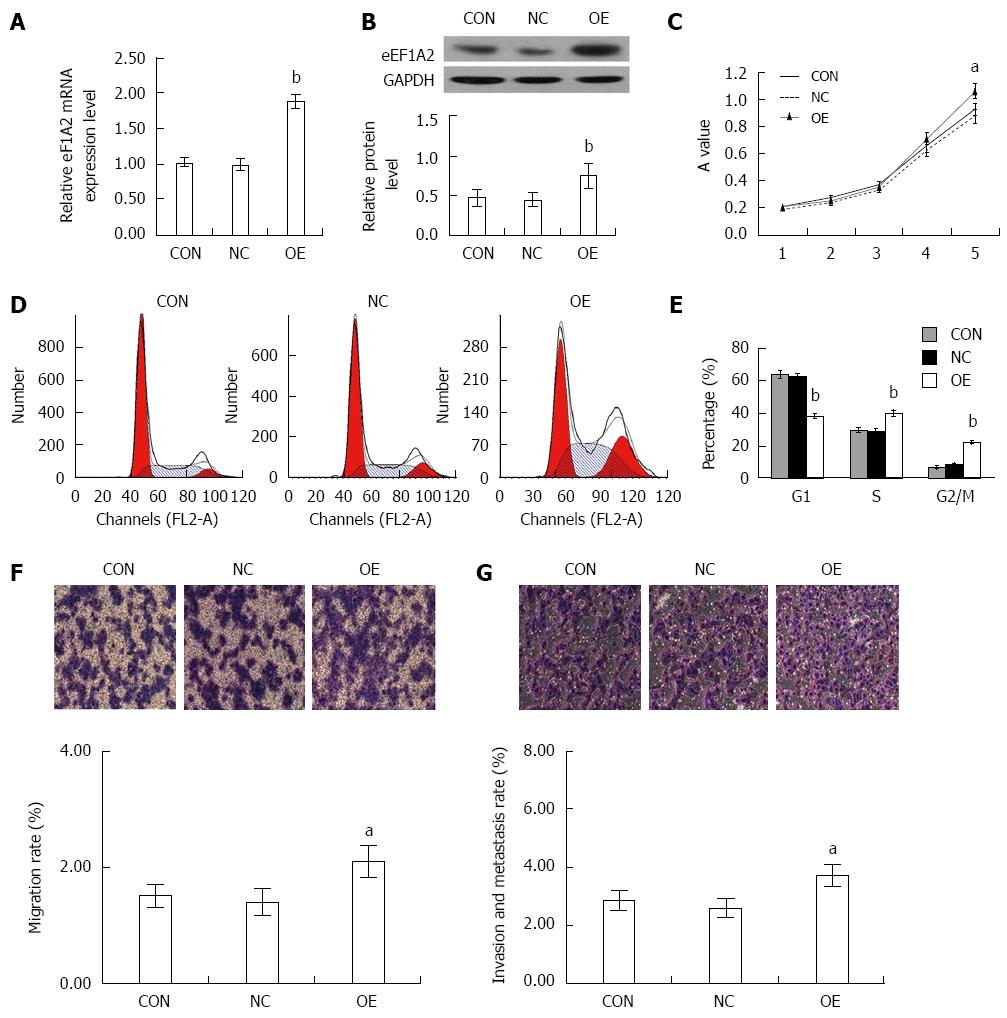
Figure 8 Overexpression of eEF1A2 promotes the proliferation, migration and invasion of SK-HEP-1 cells.
SK-HEP-1 cells were infected with the negative lentivirus (NC), without lentivirus transfection (CON), or lentivirus-based eEF1A2 overexpression (OE). A: qRT–PCR; B: Western blot analysis confirmed the expression of eEF1A2 in the SK-HEP-1 cells; C: eEF1A2 promoted the proliferation of SK-HEP-1 cells in MTT assay; D: Cell cycle distribution detected by DNA ploidy analysis; E): Percentages of SK-HEP-1 cells in the G0/G1, S and G2/M phases; F: Migration; G: Metastasis in a Transwell chamber invasion assay. Data are mean ± SD (n = 3). aP < 0.05, bP < 0.01 vs NC and CON.
- Citation: Qiu FN, Huang Y, Chen DY, Li F, Wu YA, Wu WB, Huang XL. Eukaryotic elongation factor-1α 2 knockdown inhibits hepatocarcinogenesis by suppressing PI3K/Akt/NF-κB signaling. World J Gastroenterol 2016; 22(16): 4226-4237
- URL: https://www.wjgnet.com/1007-9327/full/v22/i16/4226.htm
- DOI: https://dx.doi.org/10.3748/wjg.v22.i16.4226


