Copyright
©The Author(s) 2016.
World J Gastroenterol. Apr 28, 2016; 22(16): 4120-4135
Published online Apr 28, 2016. doi: 10.3748/wjg.v22.i16.4120
Published online Apr 28, 2016. doi: 10.3748/wjg.v22.i16.4120
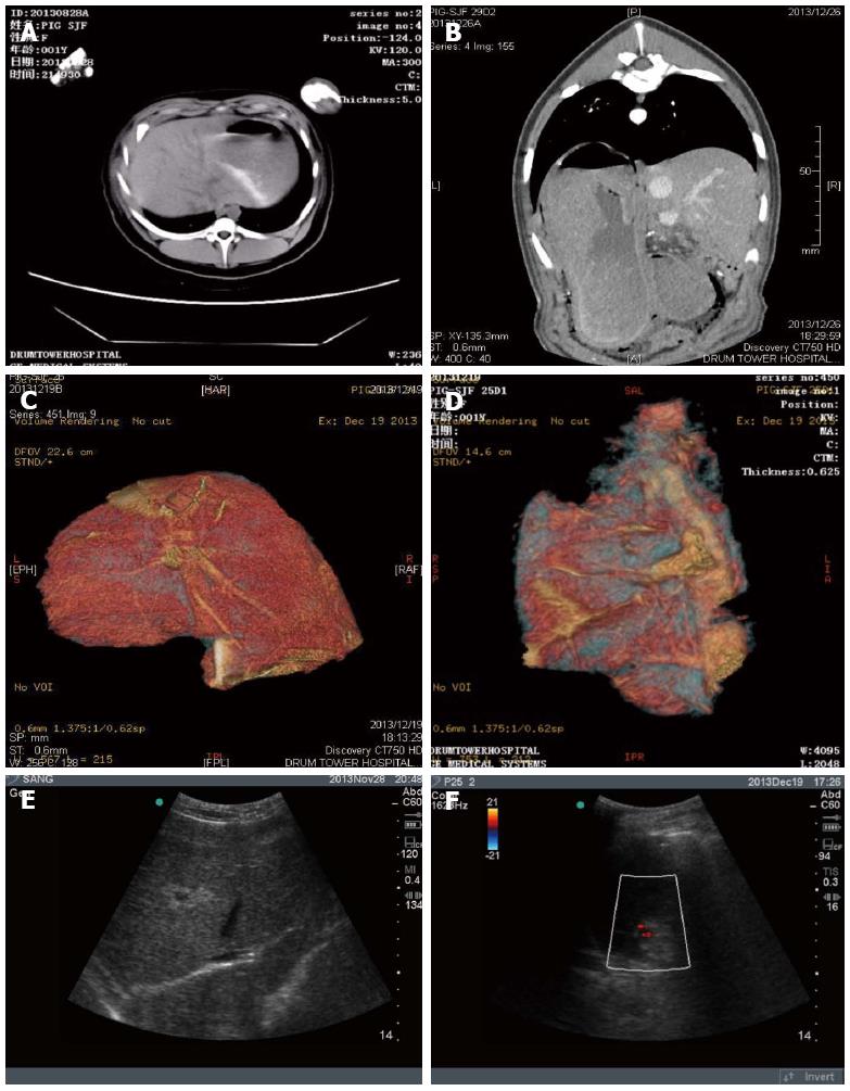
Figure 2 Imaging evaluation of residual liver volume after hepatectomy.
A and B: CT examination; C and D: CT reconstruction analysis indicated that an average 85% of liver was resected; E and F: B-scan ultrasound was performed to examine ascites (A, C, E: Sham-operated swine; B, D, F: Hepatectomized swine).
- Citation: Sang JF, Shi XL, Han B, Huang X, Huang T, Ren HZ, Ding YT. Combined mesenchymal stem cell transplantation and interleukin-1 receptor antagonism after partial hepatectomy. World J Gastroenterol 2016; 22(16): 4120-4135
- URL: https://www.wjgnet.com/1007-9327/full/v22/i16/4120.htm
- DOI: https://dx.doi.org/10.3748/wjg.v22.i16.4120


