Copyright
©The Author(s) 2016.
World J Gastroenterol. Apr 28, 2016; 22(16): 4120-4135
Published online Apr 28, 2016. doi: 10.3748/wjg.v22.i16.4120
Published online Apr 28, 2016. doi: 10.3748/wjg.v22.i16.4120
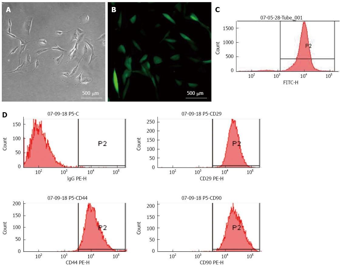
Figure 1 Characterization of mesenchymal stem cells (MSCs).
MSCs transfected with a lentiviral vector encoding green fluorescent protein (GFP) were cultured for 3 d in vitro and observed by A: Light and B: Fluorescent microscopy (magnification × 200, scale bar = 500 μm); C: Flow cytometry of MSCs revealed > 97% expressed GFP after propagation; D: Surface markers of the cultured MSCs were identified by flow cytometry: > 90% of GFP-MSCs were positive for CD29 (upper right), CD44 (lower left) and CD90 (lower right); isotypic antibodies served as the control (upper left).
- Citation: Sang JF, Shi XL, Han B, Huang X, Huang T, Ren HZ, Ding YT. Combined mesenchymal stem cell transplantation and interleukin-1 receptor antagonism after partial hepatectomy. World J Gastroenterol 2016; 22(16): 4120-4135
- URL: https://www.wjgnet.com/1007-9327/full/v22/i16/4120.htm
- DOI: https://dx.doi.org/10.3748/wjg.v22.i16.4120


