Copyright
©The Author(s) 2016.
World J Gastroenterol. Apr 28, 2016; 22(16): 4109-4119
Published online Apr 28, 2016. doi: 10.3748/wjg.v22.i16.4109
Published online Apr 28, 2016. doi: 10.3748/wjg.v22.i16.4109
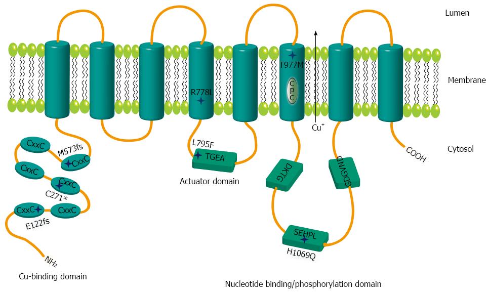
Figure 1 Schematic representation of mutations within ATP7B.
Relative positions of the mutations are shown by stars. The six metal binding domains at the N terminal are indicated by CxxC. Sequence motifs TGEA, DKTG, SEHPL, GDGVND, and CPC depict conserved elements of copper (Cu) ATPases.
- Citation: Chandhok G, Horvath J, Aggarwal A, Bhatt M, Zibert A, Schmidt HH. Functional analysis and drug response to zinc and D-penicillamine in stable ATP7B mutant hepatic cell lines. World J Gastroenterol 2016; 22(16): 4109-4119
- URL: https://www.wjgnet.com/1007-9327/full/v22/i16/4109.htm
- DOI: https://dx.doi.org/10.3748/wjg.v22.i16.4109


