Copyright
©The Author(s) 2016.
World J Gastroenterol. Apr 14, 2016; 22(14): 3837-3844
Published online Apr 14, 2016. doi: 10.3748/wjg.v22.i14.3837
Published online Apr 14, 2016. doi: 10.3748/wjg.v22.i14.3837
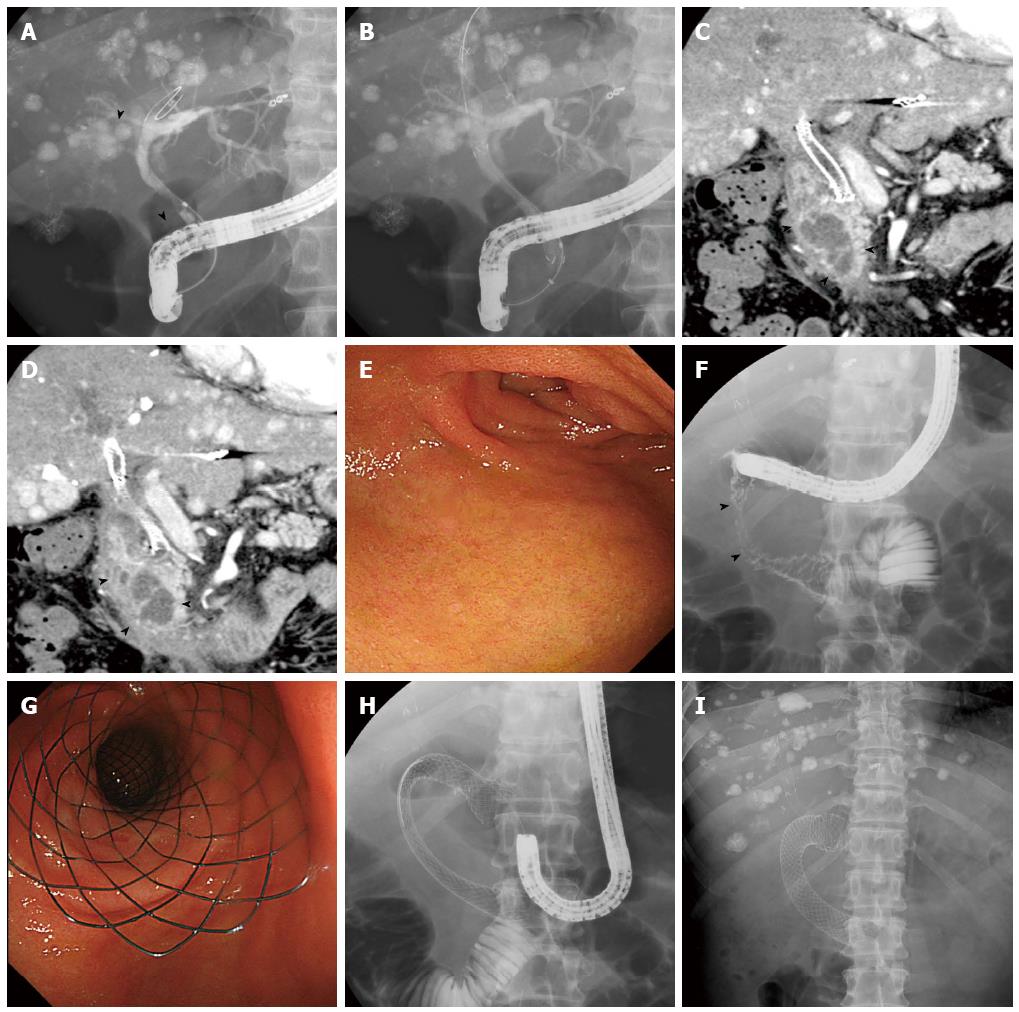
Figure 1 Case of hepatocellular carcinoma.
A: Cholangiogram showing a biliary stricture caused by primary tumor and lymph node metastasis in the right hepatic duct and middle bile duct (arrow heads); B: A self-expandable metal stent was inserted endoscopically; C, D: Coronal sections of contrast-enhanced computed tomography images show duodenal invasion of lymph node metastasis (arrow heads); E: Endoscopic view showing the oral side of the stricture at the superior duodenal angle; F: Injection of contrast material demonstrates a stricture in the second duodenal segment (arrow heads); G, H: A nitinol metal stent was placed in the shape of the character “C” from the stomach pylorus to the third duodenal segment (G: Endoscopic view; H: X-ray); I: X-ray image taken 1 wk later shows sufficient expansion and stability of the duodenal stent.
- Citation: Sasaki R, Sakai Y, Tsuyuguchi T, Nishikawa T, Fujimoto T, Mikami S, Sugiyama H, Yokosuka O. Endoscopic management of unresectable malignant gastroduodenal obstruction with a nitinol uncovered metal stent: A prospective Japanese multicenter study. World J Gastroenterol 2016; 22(14): 3837-3844
- URL: https://www.wjgnet.com/1007-9327/full/v22/i14/3837.htm
- DOI: https://dx.doi.org/10.3748/wjg.v22.i14.3837


