Copyright
©The Author(s) 2016.
World J Gastroenterol. Apr 7, 2016; 22(13): 3693-3700
Published online Apr 7, 2016. doi: 10.3748/wjg.v22.i13.3693
Published online Apr 7, 2016. doi: 10.3748/wjg.v22.i13.3693
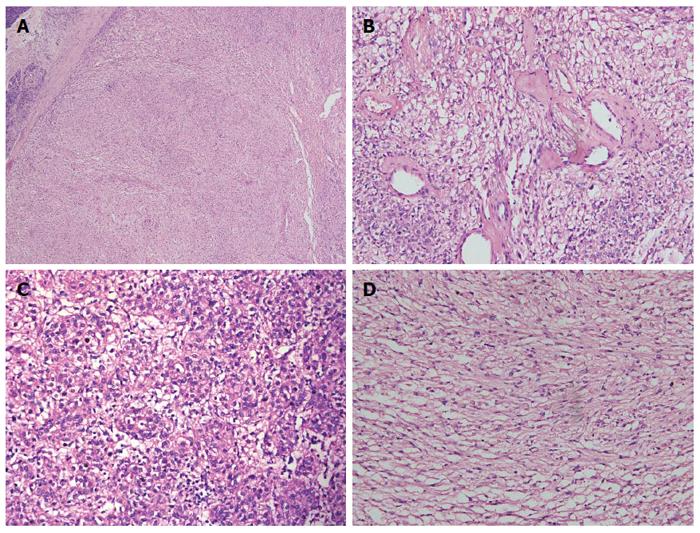
Figure 5 Microscopic examination.
A: Microscopy showing clear boundaries between the tumor and adjacent pancreatic tissue; B: Mounts of vessel were in mesenchyma, with hyaline degeneration present; C: Epithelioid tumor cell; D: Spindle-shaped tumor cell with bright or slightly eosinophilic granule. A, B, C and D: Hematoxylin-eosin staining.
- Citation: Jiang H, Ta N, Huang XY, Zhang MH, Xu JJ, Zheng KL, Jin G, Zheng JM. Pancreatic perivascular epithelioid cell tumor: A case report with clinicopathological features and a literature review. World J Gastroenterol 2016; 22(13): 3693-3700
- URL: https://www.wjgnet.com/1007-9327/full/v22/i13/3693.htm
- DOI: https://dx.doi.org/10.3748/wjg.v22.i13.3693


