Copyright
©The Author(s) 2016.
World J Gastroenterol. Mar 28, 2016; 22(12): 3355-3362
Published online Mar 28, 2016. doi: 10.3748/wjg.v22.i12.3355
Published online Mar 28, 2016. doi: 10.3748/wjg.v22.i12.3355
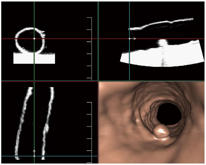
Figure 2 The 3D endoluminal reformatted image of ultrasound virtual endoscopy clearly depicting an 8-mm polyp.
We could observe the polyp on the 2D transversal, sagittal and coronal ultrasonography simultaneously.
- Citation: Liu JY, Chen LD, Cai HS, Liang JY, Xu M, Huang Y, Li W, Feng ST, Xie XY, Lu MD, Wang W. Ultrasound virtual endoscopy: Polyp detection and reliability of measurement in an in vitro study with pig intestine specimens. World J Gastroenterol 2016; 22(12): 3355-3362
- URL: https://www.wjgnet.com/1007-9327/full/v22/i12/3355.htm
- DOI: https://dx.doi.org/10.3748/wjg.v22.i12.3355


