Copyright
©The Author(s) 2016.
World J Gastroenterol. Mar 28, 2016; 22(12): 3355-3362
Published online Mar 28, 2016. doi: 10.3748/wjg.v22.i12.3355
Published online Mar 28, 2016. doi: 10.3748/wjg.v22.i12.3355
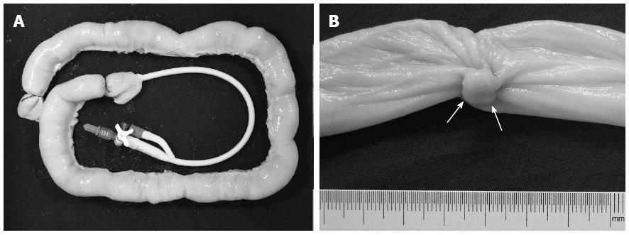
Figure 1 A pig intestine specimen and simulated polyps.
A: A porcine intestine specimen with a flexure configuration was placed on a black sponge. One end was double-tied with sutures. A catheter used for intestinal distention during the USVE and CTC was placed through the other end. Optical colonoscopy was also performed through the latter end after removal of the catheter; B: A simulated polyp with a maximum diameter of 8 mm (arrows) was sutured to the inverted intestine specimen. USVE: Ultrasound virtual endoscopy; CTC: Computed tomography colonography.
- Citation: Liu JY, Chen LD, Cai HS, Liang JY, Xu M, Huang Y, Li W, Feng ST, Xie XY, Lu MD, Wang W. Ultrasound virtual endoscopy: Polyp detection and reliability of measurement in an in vitro study with pig intestine specimens. World J Gastroenterol 2016; 22(12): 3355-3362
- URL: https://www.wjgnet.com/1007-9327/full/v22/i12/3355.htm
- DOI: https://dx.doi.org/10.3748/wjg.v22.i12.3355


