Copyright
©The Author(s) 2016.
World J Gastroenterol. Mar 21, 2016; 22(11): 3296-3301
Published online Mar 21, 2016. doi: 10.3748/wjg.v22.i11.3296
Published online Mar 21, 2016. doi: 10.3748/wjg.v22.i11.3296
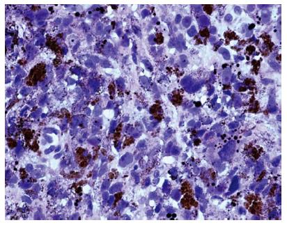
Figure 5 Histological examination showed that the excised tumor tissue was composed of non-organized and pleomorphic cells exhibiting atypical nuclei, and abundant melanin granules.
Hematoxylin-eosin staining, magnification × 400.
- Citation: Wang L, Zong L, Nakazato H, Wang WY, Li CF, Shi YF, Zhang GC, Tang T. Primary advanced esophago-gastric melanoma: A rare case. World J Gastroenterol 2016; 22(11): 3296-3301
- URL: https://www.wjgnet.com/1007-9327/full/v22/i11/3296.htm
- DOI: https://dx.doi.org/10.3748/wjg.v22.i11.3296


