Copyright
©The Author(s) 2016.
World J Gastroenterol. Mar 21, 2016; 22(11): 3242-3251
Published online Mar 21, 2016. doi: 10.3748/wjg.v22.i11.3242
Published online Mar 21, 2016. doi: 10.3748/wjg.v22.i11.3242
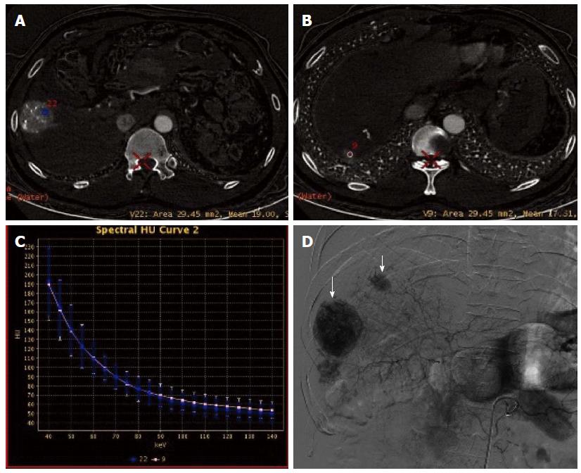
Figure 7 Same case as in Figure 6.
A: Placement of a region of interest in the primary lesion; B: Placement of a region of interest in the lesion in the right lobe; C: The spectral curves for the two lesions were roughly same, suggesting tumor homogeneity; D: Digital subtraction angiography confirmed multiple tumor stains (arrow).
- Citation: Liu QY, He CD, Zhou Y, Huang D, Lin H, Wang Z, Wang D, Wang JQ, Liao LP. Application of gemstone spectral imaging for efficacy evaluation in hepatocellular carcinoma after transarterial chemoembolization. World J Gastroenterol 2016; 22(11): 3242-3251
- URL: https://www.wjgnet.com/1007-9327/full/v22/i11/3242.htm
- DOI: https://dx.doi.org/10.3748/wjg.v22.i11.3242


