Copyright
©The Author(s) 2016.
World J Gastroenterol. Mar 21, 2016; 22(11): 3242-3251
Published online Mar 21, 2016. doi: 10.3748/wjg.v22.i11.3242
Published online Mar 21, 2016. doi: 10.3748/wjg.v22.i11.3242
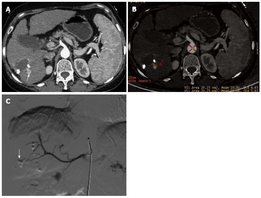
Figure 4 Recurrence of hepatocellular cancer in the right lobe after transcatheter arterial chemoembolization.
A: Sixty-eight keV monochromatic image showed obvious enlargement and enhancement of recurrent lesion in tumor edge (arrow); B: The effective iodine content of the recurrent lesion (29.89 ± 5.83) was significantly higher than that for normal liver parenchyma (10.0 ± 4.45), suggesting obvious enhancement and entry of contrast medium to the lesion; C: Digital subtraction angiography confirmed tumor stain in the recurrent lesion in parenchymal phase (arrow).
- Citation: Liu QY, He CD, Zhou Y, Huang D, Lin H, Wang Z, Wang D, Wang JQ, Liao LP. Application of gemstone spectral imaging for efficacy evaluation in hepatocellular carcinoma after transarterial chemoembolization. World J Gastroenterol 2016; 22(11): 3242-3251
- URL: https://www.wjgnet.com/1007-9327/full/v22/i11/3242.htm
- DOI: https://dx.doi.org/10.3748/wjg.v22.i11.3242


