Copyright
©The Author(s) 2016.
World J Gastroenterol. Mar 21, 2016; 22(11): 3242-3251
Published online Mar 21, 2016. doi: 10.3748/wjg.v22.i11.3242
Published online Mar 21, 2016. doi: 10.3748/wjg.v22.i11.3242
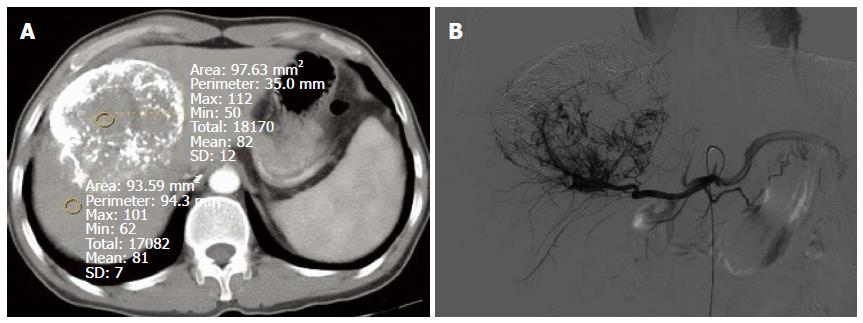
Figure 2 Giant hepatocellular cancer in the lateral borders of the left and right liver lobes after transcatheter arterial chemoembolization.
A: The computed tomography (CT) value of the defect of iodine deposition was comparable to that of normal liver parenchyma in conventional CT image, and there was no obvious enhancement; B: Digital subtraction angiography revealed enlarged tumor blood vessels and tumor stain in the defect of iodine deposition.
- Citation: Liu QY, He CD, Zhou Y, Huang D, Lin H, Wang Z, Wang D, Wang JQ, Liao LP. Application of gemstone spectral imaging for efficacy evaluation in hepatocellular carcinoma after transarterial chemoembolization. World J Gastroenterol 2016; 22(11): 3242-3251
- URL: https://www.wjgnet.com/1007-9327/full/v22/i11/3242.htm
- DOI: https://dx.doi.org/10.3748/wjg.v22.i11.3242


