Copyright
©The Author(s) 2016.
World J Gastroenterol. Mar 21, 2016; 22(11): 3242-3251
Published online Mar 21, 2016. doi: 10.3748/wjg.v22.i11.3242
Published online Mar 21, 2016. doi: 10.3748/wjg.v22.i11.3242
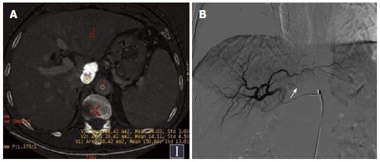
Figure 1 Hepatocellular cancer in the caudate lobe after transcatheter arterial chemoembolization.
A: Iodine (water) based image. The effective iodine content of the defect of iodine deposition (154.10 ± 8.07) was significantly higher than that for normal liver parenchyma and the aorta (67.36 ± 4.87), revealing no residual tumor; B: Digital subtraction angiography image revealed no tumor vessels or stain, and obvious iodized oil deposition was visible (arrow).
- Citation: Liu QY, He CD, Zhou Y, Huang D, Lin H, Wang Z, Wang D, Wang JQ, Liao LP. Application of gemstone spectral imaging for efficacy evaluation in hepatocellular carcinoma after transarterial chemoembolization. World J Gastroenterol 2016; 22(11): 3242-3251
- URL: https://www.wjgnet.com/1007-9327/full/v22/i11/3242.htm
- DOI: https://dx.doi.org/10.3748/wjg.v22.i11.3242


