Copyright
©The Author(s) 2016.
World J Gastroenterol. Mar 14, 2016; 22(10): 2971-2980
Published online Mar 14, 2016. doi: 10.3748/wjg.v22.i10.2971
Published online Mar 14, 2016. doi: 10.3748/wjg.v22.i10.2971
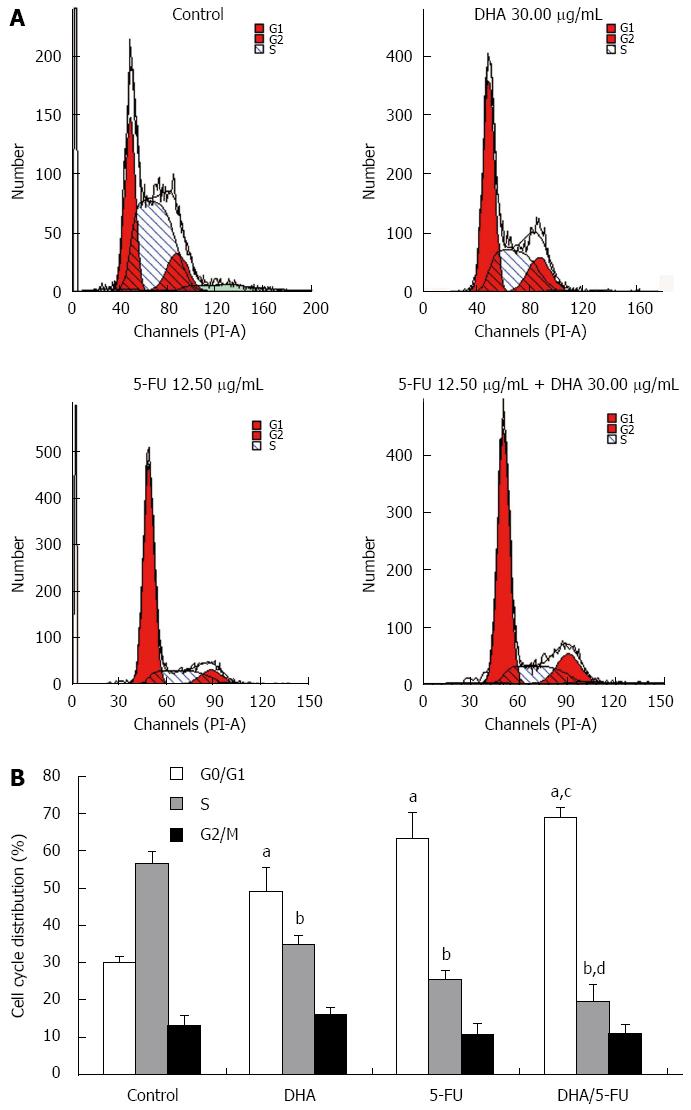
Figure 3 Cell cycle analysis of AGS cell after docosahexaenoic acid or 5-fluorouracil treatment or DHA/5-FU co-administration.
Docosahexaenoic acid (DHA) enhanced 5-fluorouracil (5-FU)-induced cell cycle arrest at G0/G1 phase in AGS cells. AGS cells were exposed to medium only (control), DHA alone (30.00 μg/mL), 5-FU alone (12.5 μg/mL), or DHA (30.00 μg/mL) plus 5-FU (12.50 μg/mL) for 48 h. A: A representative set of results showing cell cycle distribution in different groups; B: An accumulation of cells in the G0/G1 phase and a decrease of cells in the S phase after treatment with DHA or 5-FU, and a significant potentiation in accumulation of cells in G0/G1 phase and reduction in S phase after the combined treatment of 5-FU with DHA. Data shown are a representative example of three separate experiments with similar results. aP < 0.05 vs control cells in the G0/G1 phase; bP < 0.05 vs control cells in the S phase; cP < 0.05 vs cells treated with DHA or 5-FU in the G0/G1 phase; dP < 0.05 vs cells treated with DHA or 5-FU in the S phase.
- Citation: Gao K, Liang Q, Zhao ZH, Li YF, Wang SF. Synergistic anticancer properties of docosahexaenoic acid and 5-fluorouracil through interference with energy metabolism and cell cycle arrest in human gastric cancer cell line AGS cells. World J Gastroenterol 2016; 22(10): 2971-2980
- URL: https://www.wjgnet.com/1007-9327/full/v22/i10/2971.htm
- DOI: https://dx.doi.org/10.3748/wjg.v22.i10.2971


