Copyright
©The Author(s) 2016.
World J Gastroenterol. Mar 14, 2016; 22(10): 2971-2980
Published online Mar 14, 2016. doi: 10.3748/wjg.v22.i10.2971
Published online Mar 14, 2016. doi: 10.3748/wjg.v22.i10.2971
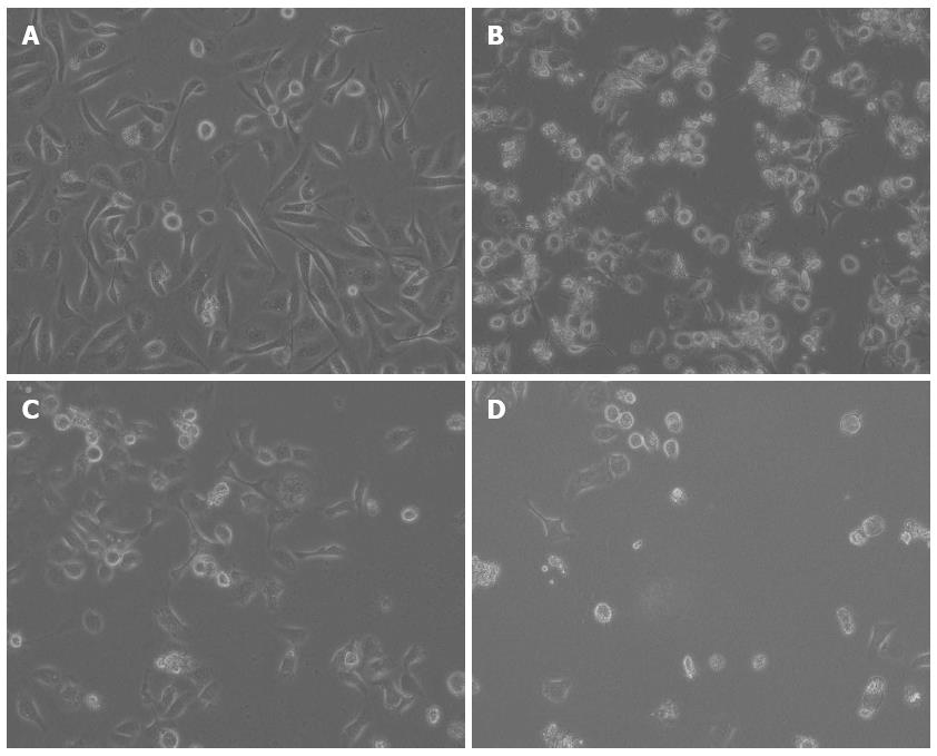
Figure 2 Morphological changes in AGS cells treated with docosahexaenoic acid and 5-fluorouracil alone or in combination.
AGS cells were treated with medium only (control, A), docosahexaenoic acid (DHA) alone (30.00 μg/mL, B), 5-fluorouracil (5-FU) alone (12.50 μg/mL, C), or DHA plus 5-FU (30.00 μg/mL + 12.50 μg/mL, D) for 48 h. The cells were observed under an inverted-phase microscope. The photographs were taken at magnification × 200.
- Citation: Gao K, Liang Q, Zhao ZH, Li YF, Wang SF. Synergistic anticancer properties of docosahexaenoic acid and 5-fluorouracil through interference with energy metabolism and cell cycle arrest in human gastric cancer cell line AGS cells. World J Gastroenterol 2016; 22(10): 2971-2980
- URL: https://www.wjgnet.com/1007-9327/full/v22/i10/2971.htm
- DOI: https://dx.doi.org/10.3748/wjg.v22.i10.2971


