Copyright
©The Author(s) 2016.
World J Gastroenterol. Jan 7, 2016; 22(1): 89-102
Published online Jan 7, 2016. doi: 10.3748/wjg.v22.i1.89
Published online Jan 7, 2016. doi: 10.3748/wjg.v22.i1.89
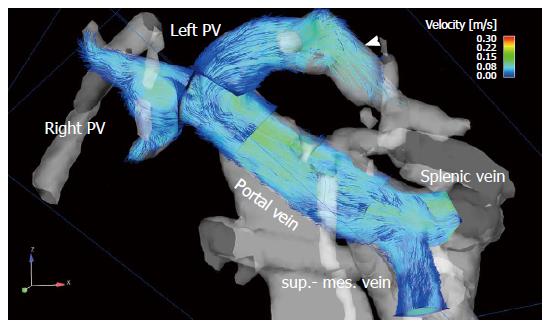
Figure 2 The portal venous system visualized by particle traces in a 68-year-old male patient with liver cirrhosis (Child-Pugh stage B).
Blue emitter planes were positioned in the splenic vein and superior mesenteric vein (sup.-mes. vein), portal vein, right (right PV) and left (left PV) intrahepatic portal vein branch. Time-resolved particle traces illustrate physiological flow in the extrahepatic portal venous system with inflow in the portal vein from the splenic and superior mesenteric veins. Flow over the left branch of the intrahepatic portal vein into a re-opened umbilical vein is visible (arrowhead).
- Citation: Stankovic Z. Four-dimensional flow magnetic resonance imaging in cirrhosis. World J Gastroenterol 2016; 22(1): 89-102
- URL: https://www.wjgnet.com/1007-9327/full/v22/i1/89.htm
- DOI: https://dx.doi.org/10.3748/wjg.v22.i1.89


