Copyright
©The Author(s) 2016.
World J Gastroenterol. Jan 7, 2016; 22(1): 284-299
Published online Jan 7, 2016. doi: 10.3748/wjg.v22.i1.284
Published online Jan 7, 2016. doi: 10.3748/wjg.v22.i1.284
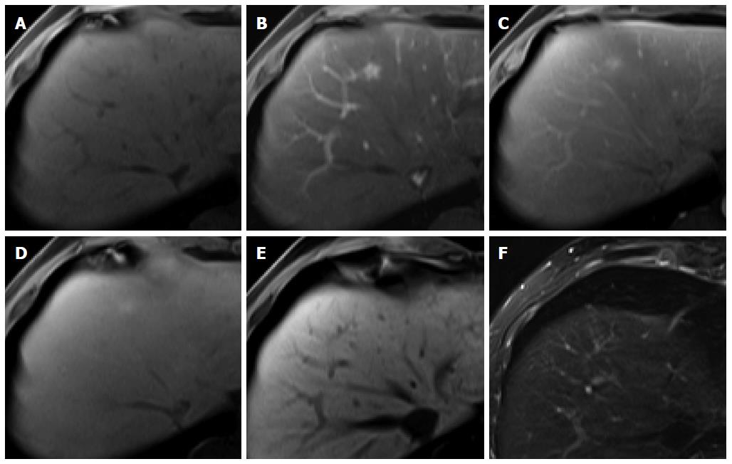
Figure 5 Focal nodular hyperplasia-like nodule in a 45-year-old man with hepatitis B infection.
A: Precontrast T1-weighted image shows isointensity of the tumor; B: Hepatic arterial phase using gadoxetic acid shows lobulating-contoured, marked enhanced nodule in segment 4; C, D: Portal venous and transitional phases show slight hyperenhancement of the tumor relative to the liver parenchyma; E: Hepatobiliary phase shows isointensity or subtle peripheral ring-like enhancement of the tumor; F: T2-weighted image shows isointensity of the tumor.
- Citation: Park YS, Lee CH, Kim JW, Shin S, Park CM. Differentiation of hepatocellular carcinoma from its various mimickers in liver magnetic resonance imaging: What are the tips when using hepatocyte-specific agents? World J Gastroenterol 2016; 22(1): 284-299
- URL: https://www.wjgnet.com/1007-9327/full/v22/i1/284.htm
- DOI: https://dx.doi.org/10.3748/wjg.v22.i1.284


