Copyright
©The Author(s) 2016.
World J Gastroenterol. Jan 7, 2016; 22(1): 221-231
Published online Jan 7, 2016. doi: 10.3748/wjg.v22.i1.221
Published online Jan 7, 2016. doi: 10.3748/wjg.v22.i1.221
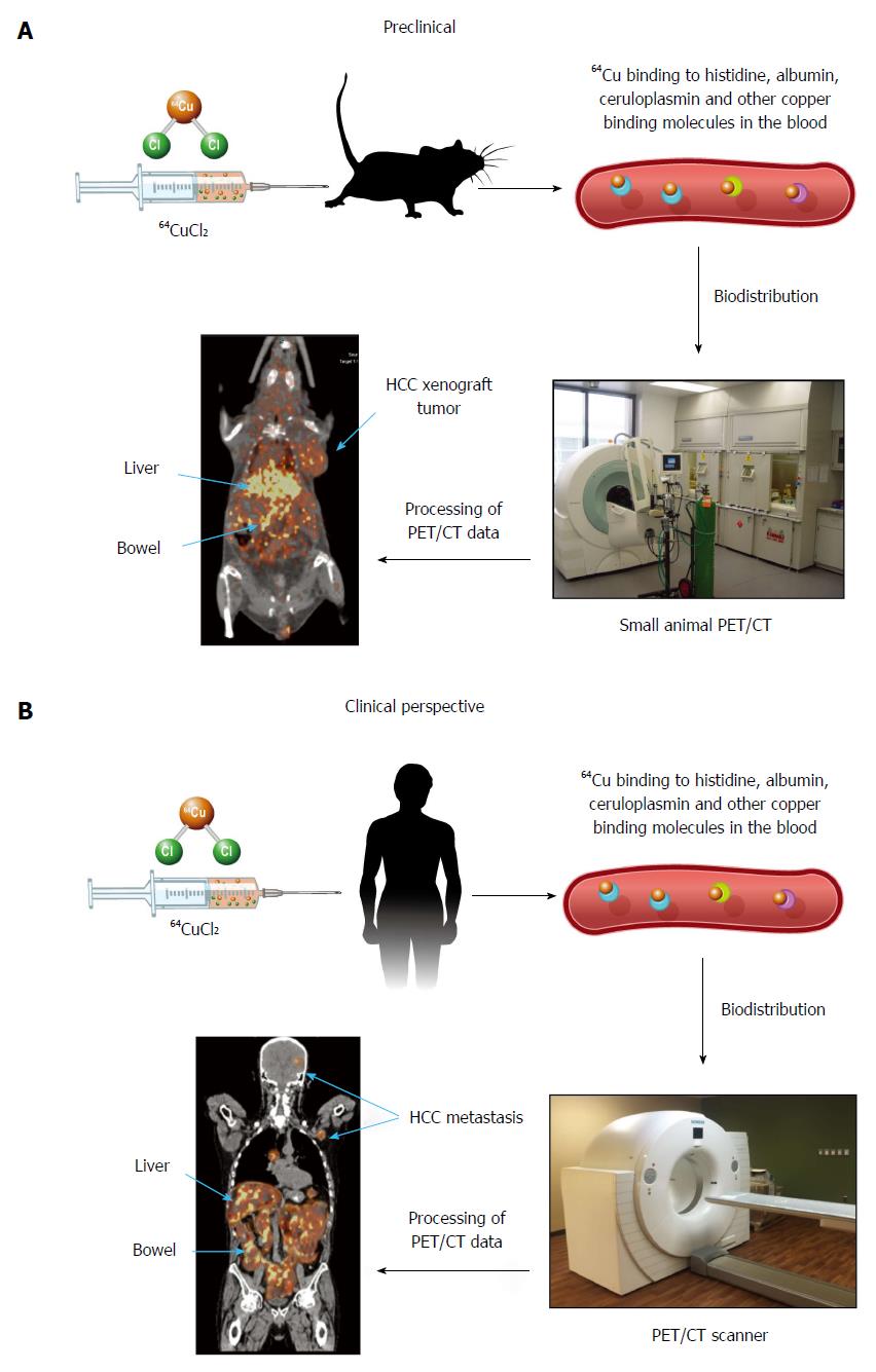
Figure 1 Metabolic imaging of metastasis of hepatocellular carcinoma with 64CuCl2-position emission tomography and computed tomography.
A: Preclinical metabolic imaging of HCC xenografts in mice injected with 64CuCl2 as a tracer. 64Cu bound to copper binding molecules in the blood immediately after intravenous injection of 64CuCl2. PET/CT images were then obtained that show the expected biodistribution of 64CuCl2 in the liver and intestinal tracts, with low uptake in the brain and muscle tissues. The HCC xenografts implanted on the shoulder showed increased 64Cu uptake on PET/CT images; B: Schematic showing the clinical perspective of metabolic imaging of HCC in humans. Human patients may be injected with 64CuCl2 and subjected to PET/CT for detection of HCC metastasis in areas of low physiological 64Cu uptake, such as brain and musculoskeletal tissues. HCC: Hepatocellular carcinoma; PET/CT: Hybrid positron emission tomography and computed tomography.
- Citation: Wachsmann J, Peng F. Molecular imaging and therapy targeting copper metabolism in hepatocellular carcinoma. World J Gastroenterol 2016; 22(1): 221-231
- URL: https://www.wjgnet.com/1007-9327/full/v22/i1/221.htm
- DOI: https://dx.doi.org/10.3748/wjg.v22.i1.221


