Copyright
©The Author(s) 2016.
World J Gastroenterol. Jan 7, 2016; 22(1): 205-220
Published online Jan 7, 2016. doi: 10.3748/wjg.v22.i1.205
Published online Jan 7, 2016. doi: 10.3748/wjg.v22.i1.205
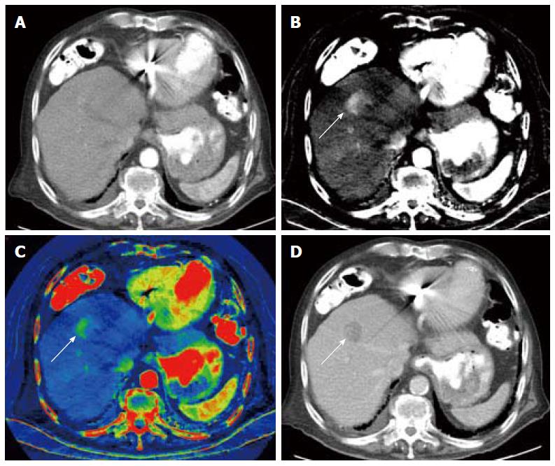
Figure 14 Dual-energy computed tomography of hepatocellular carcinoma.
Nodular outline is suggestive of cirrhosis, arterial phase single energy computed tomography (SECT) image acquired at 140kVp demonstrates a vague focus of arterial enhancement that is difficult to differentiate from surrounding liver parenchyma (A), arterial phase DECT material decomposition iodine (MD-I) image shows uptake of iodine independently from inherent tissue attenuation, clearly demonstrating a nodular hyperenhancing lesion (B), arterial phase color overlay MD-I image also depicts the lesion well (C). Portal venous phase SECT image acquired at 120 kVp demonstrates characteristic wash-out (D). MD-I images improve detection and characterization of this small hepatocellular carcinoma. (Courtesy of Drs. Andrea Prochowski and Dushyant Sahani, Massachusetts General Hospital, Boston, MA, United States).
- Citation: Hennedige T, Venkatesh SK. Advances in computed tomography and magnetic resonance imaging of hepatocellular carcinoma. World J Gastroenterol 2016; 22(1): 205-220
- URL: https://www.wjgnet.com/1007-9327/full/v22/i1/205.htm
- DOI: https://dx.doi.org/10.3748/wjg.v22.i1.205


