Copyright
©The Author(s) 2016.
World J Gastroenterol. Jan 7, 2016; 22(1): 205-220
Published online Jan 7, 2016. doi: 10.3748/wjg.v22.i1.205
Published online Jan 7, 2016. doi: 10.3748/wjg.v22.i1.205
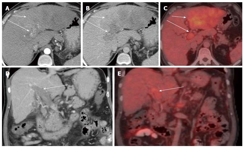
Figure 10 Vascular invasion.
A large ill-defined left lobe mass with no significant arterial enhancement (A) and washout in the portal venous phase (B). An FDG-PET CT was done which revealed uptake in the left lobe mass (C) consistent with a hypermetabolic tumour. Arterial enhancement is noted within the distended thrombus filled portal veins in (A) with subsequent washout (B) suggestive of tumour thrombus. The tumour thrombus also demonstrates increased uptake on FDG-PET (C). Coronal images better depict the distended thrombus filled portal vein (D) with increased uptake on PET/CT (E) (short arrow: tumour; long arrow: tumour thrombus). PET: Positron emission tomography; CT: Computed tomography; FDG: Fluoro-2-deoxy-D-glucose.
- Citation: Hennedige T, Venkatesh SK. Advances in computed tomography and magnetic resonance imaging of hepatocellular carcinoma. World J Gastroenterol 2016; 22(1): 205-220
- URL: https://www.wjgnet.com/1007-9327/full/v22/i1/205.htm
- DOI: https://dx.doi.org/10.3748/wjg.v22.i1.205


