Copyright
©The Author(s) 2016.
World J Gastroenterol. Jan 7, 2016; 22(1): 205-220
Published online Jan 7, 2016. doi: 10.3748/wjg.v22.i1.205
Published online Jan 7, 2016. doi: 10.3748/wjg.v22.i1.205
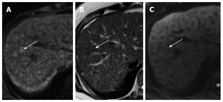
Figure 4 Dysplastic nodules may appears hyperintense on T1W (A), iso-hypointense on T2W (B) but do not show restricted diffusion (C).
- Citation: Hennedige T, Venkatesh SK. Advances in computed tomography and magnetic resonance imaging of hepatocellular carcinoma. World J Gastroenterol 2016; 22(1): 205-220
- URL: https://www.wjgnet.com/1007-9327/full/v22/i1/205.htm
- DOI: https://dx.doi.org/10.3748/wjg.v22.i1.205


