Copyright
©The Author(s) 2016.
World J Gastroenterol. Jan 7, 2016; 22(1): 205-220
Published online Jan 7, 2016. doi: 10.3748/wjg.v22.i1.205
Published online Jan 7, 2016. doi: 10.3748/wjg.v22.i1.205
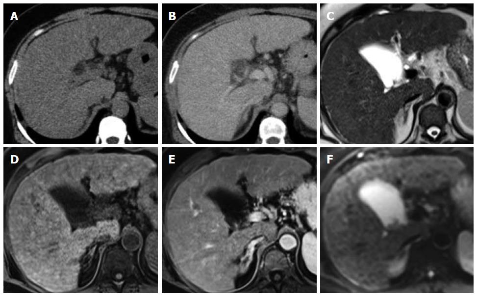
Figure 2 The liver demonstrates a nodular outline consistent with cirrhosis and multiple small regenerative nodules that are isodense on unenhanced (A) and portal venous phase (B) on computed tomography, predominantly isointense on T2W (C) and T1W (D) sequences with no evidence of arterial enhancement (E) or restricted diffusion (F).
- Citation: Hennedige T, Venkatesh SK. Advances in computed tomography and magnetic resonance imaging of hepatocellular carcinoma. World J Gastroenterol 2016; 22(1): 205-220
- URL: https://www.wjgnet.com/1007-9327/full/v22/i1/205.htm
- DOI: https://dx.doi.org/10.3748/wjg.v22.i1.205


