Copyright
©The Author(s) 2016.
World J Gastroenterol. Jan 7, 2016; 22(1): 103-111
Published online Jan 7, 2016. doi: 10.3748/wjg.v22.i1.103
Published online Jan 7, 2016. doi: 10.3748/wjg.v22.i1.103
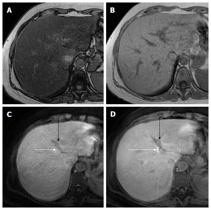
Figure 5 Reduced hepatobiliary phase enhancement due to severe hepatic steatosis in a 42-year-old woman with hepatitis C virus-related chronic hepatitis.
A, B: T1-weighted gradient-echo images show diffuse signal intensity decrease of the liver on out-of-phase (A) image compared with that on the in-phase image (B), indicating severe hepatic steatosis; C: On 10 min hepatobiliary phase, gadoxetic acid enhanced magnetic resonance imaging, left portal vein (black arrow) shows iso- to hypointensity to liver parenchyma; D: On 20 min hepatobiliary phase left portal vein shows slight hypointensity to liver parenchyma. Enhancement of bile ducts (white arrows) is less intense on 10 min hepatobiliary phase than that on 20 min hepatobiliary phase, indicating delayed biliary elimination of gadoxetic acid.
- Citation: Agnello F, Dioguardi Burgio M, Picone D, Vernuccio F, Cabibbo G, Giannitrapani L, Taibbi A, Agrusa A, Bartolotta TV, Galia M, Lagalla R, Midiri M, Brancatelli G. Magnetic resonance imaging of the cirrhotic liver in the era of gadoxetic acid. World J Gastroenterol 2016; 22(1): 103-111
- URL: https://www.wjgnet.com/1007-9327/full/v22/i1/103.htm
- DOI: https://dx.doi.org/10.3748/wjg.v22.i1.103


