Copyright
©The Author(s) 2016.
World J Gastroenterol. Jan 7, 2016; 22(1): 103-111
Published online Jan 7, 2016. doi: 10.3748/wjg.v22.i1.103
Published online Jan 7, 2016. doi: 10.3748/wjg.v22.i1.103
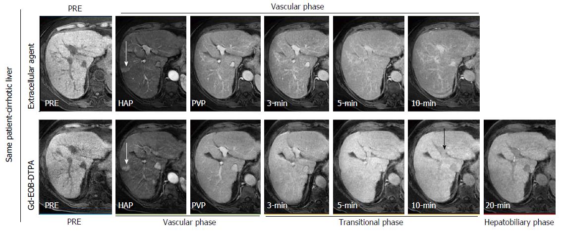
Figure 2 Intraindividual differences in hepatic enhancement in cirrhotic liver between extra-cellular contrast agent (top row) and gadoxetic acid (bottom row) in a 69-year-old woman with hepatitis C virus-related cirrhosis.
On contrast-enhanced magnetic resonance (MR) images obtained with an extra-cellular agent liver enhancement peaks on portal venous phase and then slightly decreases. On contrast-enhanced MR images obtained with gadoxetic acid, liver enhancement shows a stepwise increase from the hepatic arterial phase to the 20 min phase. Vascular enhancement is more prolonged with extra-cellular agent than with gadoxetic acid, indicating a slower vascular elimination. On 10 min, the intrahepatic vessels (black arrow) show slight hypointensity to the liver, and the bile ducts are not opacified. These findings indicate hepatic dysfunction and a prolonged transitional phase. Also, note a wedge shaped enhancing area in the hepatic arterial phase (white arrow), with lack of washout on portal venous phase and isointensity on hepatobiliary phase, due to arterioportal shunt. PRE: Precontrast; HAP: Late hepatic arterial phase; PVP: Portal venous phase.
- Citation: Agnello F, Dioguardi Burgio M, Picone D, Vernuccio F, Cabibbo G, Giannitrapani L, Taibbi A, Agrusa A, Bartolotta TV, Galia M, Lagalla R, Midiri M, Brancatelli G. Magnetic resonance imaging of the cirrhotic liver in the era of gadoxetic acid. World J Gastroenterol 2016; 22(1): 103-111
- URL: https://www.wjgnet.com/1007-9327/full/v22/i1/103.htm
- DOI: https://dx.doi.org/10.3748/wjg.v22.i1.103


