Copyright
©The Author(s) 2015.
World J Gastroenterol. Mar 7, 2015; 21(9): 2830-2835
Published online Mar 7, 2015. doi: 10.3748/wjg.v21.i9.2830
Published online Mar 7, 2015. doi: 10.3748/wjg.v21.i9.2830
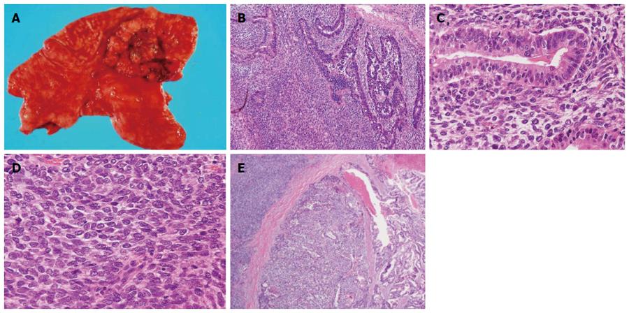
Figure 2 Histopathology.
A: Surgical resection revealed a large tumor (75 mm × 75 mm) with central ulceration (Borrmann type III) occupying the anterior wall of the upper body of the stomach; Hematoxylin and eosin staining revealed B: Biphasic structure consisting of tubular adenocarcinoma and spindle cell sarcoma (20 ×); C: Epithelial components showing that the parenchyma structure of the common gastric adenocarcinoma was surrounded with sarcomatous stroma (200 ×); D: Sarcomatous cells proliferating diffusely in a stream pattern (200 ×); E: Two transitional areas of carcinoma and sarcoma components (20 ×).
- Citation: Gohongi T, Iida H, Gunji N, Orii K, Ogata T. Postsurgical radiation therapy for gastric carcinosarcoma with c-kit expression: A case report. World J Gastroenterol 2015; 21(9): 2830-2835
- URL: https://www.wjgnet.com/1007-9327/full/v21/i9/2830.htm
- DOI: https://dx.doi.org/10.3748/wjg.v21.i9.2830


