Copyright
©The Author(s) 2015.
World J Gastroenterol. Mar 7, 2015; 21(9): 2820-2825
Published online Mar 7, 2015. doi: 10.3748/wjg.v21.i9.2820
Published online Mar 7, 2015. doi: 10.3748/wjg.v21.i9.2820
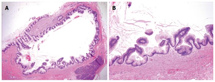
Figure 3 Pancreatic duct histology.
A: IPMN involving the main pancreatic duct (40 × magnification); B: Neoplastic cells show hyperchromatic nuclei with abundant intracytoplasmic mucin. No high-grade dysplastic cells are present (100 × magnification).
- Citation: Flanagan MR, Jayaraj A, Xiong W, Yeh MM, Raskind WH, Pillarisetty VG. Pancreatic intraductal papillary mucinous neoplasm in a patient with Lynch syndrome. World J Gastroenterol 2015; 21(9): 2820-2825
- URL: https://www.wjgnet.com/1007-9327/full/v21/i9/2820.htm
- DOI: https://dx.doi.org/10.3748/wjg.v21.i9.2820


