Copyright
©The Author(s) 2015.
World J Gastroenterol. Mar 7, 2015; 21(9): 2700-2710
Published online Mar 7, 2015. doi: 10.3748/wjg.v21.i9.2700
Published online Mar 7, 2015. doi: 10.3748/wjg.v21.i9.2700
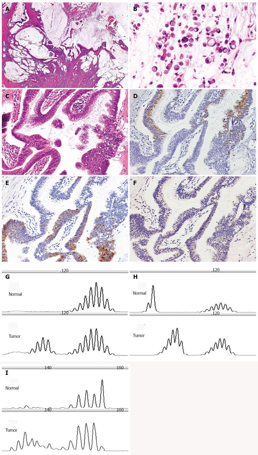
Figure 3 Small intestinal adenocarcinoma with microsatellite instability-high (case 36).
A: The tumor produced abundant mucin (HE stain, × 20); B: The tumor focally showed signet ring-like carcinoma cells (HE stain, × 200); C: The carcinomatous gland was composed of columnar cells with intracytoplasmic mucin (HE stain, × 200); D: These glands were positive for MUC2 (MUC2, × 200); E: positive for MUC5AC (MUC5AC, × 200); F: But negative for MUC6 (MUC6, × 200). In these microsatellites, the tumor was unstable. Microsatellite analysis of paired normal and tumor DNA using G: BAT25; H: BAT26; I: D17S250.
- Citation: Kumagai R, Kohashi K, Takahashi S, Yamamoto H, Hirahashi M, Taguchi K, Nishiyama K, Oda Y. Mucinous phenotype and CD10 expression of primary adenocarcinoma of the small intestine. World J Gastroenterol 2015; 21(9): 2700-2710
- URL: https://www.wjgnet.com/1007-9327/full/v21/i9/2700.htm
- DOI: https://dx.doi.org/10.3748/wjg.v21.i9.2700


