Copyright
©The Author(s) 2015.
World J Gastroenterol. Mar 7, 2015; 21(9): 2693-2699
Published online Mar 7, 2015. doi: 10.3748/wjg.v21.i9.2693
Published online Mar 7, 2015. doi: 10.3748/wjg.v21.i9.2693
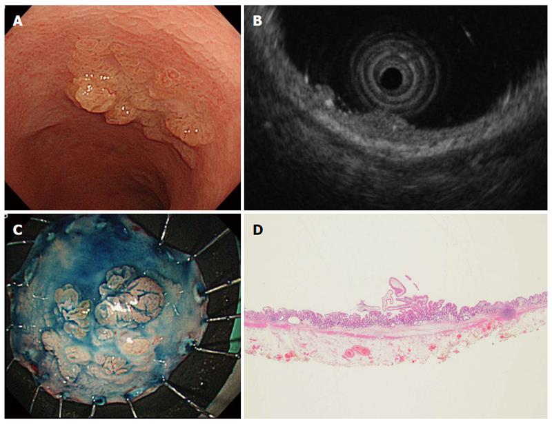
Figure 1 Lesion accurately diagnosed by endoscopic ultrasonography.
A: Colonoscopic image, showing a flat tumor with a multinodular surface in the rectum; B: On endoscopic ultrasonography, the tumor was localized to the first and second layers of the rectal wall, with no marked changes in the third layer beneath the tumor. An intramucosal lesion was diagnosed; C: Macroscopic appearance of a specimen obtained by endoscopic submucosal dissection after spraying with indigo carmine solution; D: The histopathological diagnosis was low-grade dysplasia.
- Citation: Kobayashi K, Kawagishi K, Ooka S, Yokoyama K, Sada M, Koizumi W. Clinical usefulness of endoscopic ultrasonography for the evaluation of ulcerative colitis-associated tumors. World J Gastroenterol 2015; 21(9): 2693-2699
- URL: https://www.wjgnet.com/1007-9327/full/v21/i9/2693.htm
- DOI: https://dx.doi.org/10.3748/wjg.v21.i9.2693


