Copyright
©The Author(s) 2015.
World J Gastroenterol. Feb 28, 2015; 21(8): 2542-2545
Published online Feb 28, 2015. doi: 10.3748/wjg.v21.i8.2542
Published online Feb 28, 2015. doi: 10.3748/wjg.v21.i8.2542
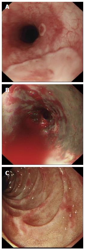
Figure 1 Endoscopic appearance.
A: Upper oesophagus showing raised whitish plaques which were thought to be viral in origin at the time of endoscopy; B: Lower oesophagus showing severe confluent oesophagitis with necrosis and active bleeding; C: Second part of duodenum showing a non specific duodenitis.
- Citation: Smith LA, Gangopadhyay M, Gaya DR. Catastrophic gastrointestinal complication of systemic immunosuppression. World J Gastroenterol 2015; 21(8): 2542-2545
- URL: https://www.wjgnet.com/1007-9327/full/v21/i8/2542.htm
- DOI: https://dx.doi.org/10.3748/wjg.v21.i8.2542


