Copyright
©The Author(s) 2015.
World J Gastroenterol. Feb 21, 2015; 21(7): 2242-2248
Published online Feb 21, 2015. doi: 10.3748/wjg.v21.i7.2242
Published online Feb 21, 2015. doi: 10.3748/wjg.v21.i7.2242
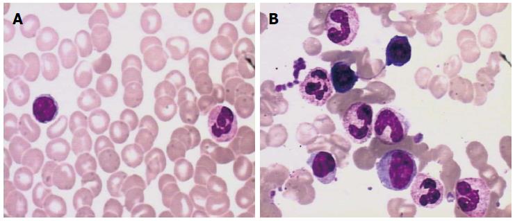
Figure 4 Representative images of blood smears and bone marrow biopsies.
The images show that the bone marrow is normal without acute myeloblastic leukemia A: Blood smear (Wright and Giemsa stain, × 1000); B: Bone marrow biopsy (Wright and Giemsa staining, × 1000).
- Citation: Huang XL, Tao J, Li JZ, Chen XL, Chen JN, Shao CK, Wu B. Gastric myeloid sarcoma without acute myeloblastic leukemia. World J Gastroenterol 2015; 21(7): 2242-2248
- URL: https://www.wjgnet.com/1007-9327/full/v21/i7/2242.htm
- DOI: https://dx.doi.org/10.3748/wjg.v21.i7.2242


